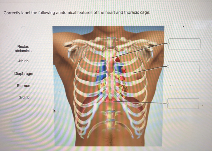
41 correctly label the following anatomical features of the heart and thoracic cage.
PDF BIO 113 LAB 1. Anatomical Terminology, Positions, Planes, and Sections ... Identify and use anatomical terms to correctly label the following regions on Figure 1: BIO 113 Fall 2011 LAB 1 Page 2 ... The heart and lungs, located in the thoracic cavity, are protected by the bony rib cage. The cavity inferior to the diaphragm is the . abdominopelvic cavity. Free Science Flashcards about ANP1040 Exam 3 - StudyStack ANP1040 Exam 3. Question. Answer. Correctly label the following anatomical features of a vertebra. Vertebral arch, Spinous Process, Nucleus Pulposus, Transvere Process, Body, Vertebral Foramen, Anulous Fibrous. Correctly identify the bones and anatomical features of the bones of the skull. Frontal Bone, Maxilla, Mandible, Zygomatic Bone ...
Thoracic cage: Anatomy and clinical notes | Kenhub The thoracic vertebrae are a group of 12 irregular bones that form the thoracic portion of the vertebral spine. According to their structure, the thoracic vertebrae can be typical and atypical. They are mostly typical vertebrae in that they are independent, have bodies, vertebral arches, and seven processes for muscular and articular connections.

Correctly label the following anatomical features of the heart and thoracic cage.
The Thoracic Cage - Anatomy and Physiology - opentextbc.ca The thoracic cage protects the heart and lungs. Thoracic Cage The thoracic cage is formed by the (a) sternum and (b) 12 pairs of ribs with their costal cartilages. The ribs are anchored posteriorly to the 12 thoracic vertebrae. The sternum consists of the manubrium, body, and xiphoid process. Solved Correctly label the following anatomical features of - Chegg 1sternum *The sternum or breastbone is a long f … View the full answer Transcribed image text: Correctly label the following anatomical features of the heart and thoracic cage. Rectus abdominis 4th rib Diaphragm INC Sternum UD OULU 3rd rib ( 1 Previous question Next question Structure of the Ribcage and Ribs - GetBodySmart 1. 2. Ribs 3-9 share many structural characteristics. In comparison, the first two ribs are shorter and more curved. Rib 1 is also flattened horizontally. The heads of ribs 1, 10, 11, and 12 have a single facet for articulation with the bodies of the thoracic vertebrae. Ribs 11 and 12 do not have necks or tubercles and the anterior tips of ...
Correctly label the following anatomical features of the heart and thoracic cage.. Anatomy And Physiology 2 Lab Exam - myilibrary.org Label each line on the pressure graph below as representing either the aorta, left atrium, or left ventricle. Identify the specific region on the graph associated with each phase of the cardiac cycle listed. Correctly label the following anatomical features of the heart and thoracic cage. Anatomy and Physiology 2 Lab exam 1 MIDTERM Flashcards - Quizlet Identify the specific region on the graph associated with each phase of the cardiac cycle listed Correctly label the following anatomical features of the heart and thoracic cage Correctly label the following vessels leading from and toward the anterior heart Correctly label the following external anatomy of the posterior heart. arteries and veins labeling quiz - Illusions By Allen The ribs protect vital organs within the thoracic cage, and they also assist with breathing. cat arteries and veins Google Search Vet tech . Detta är en online quiz som heter Label the Veins and Arteries by spoor . Labeling Arterial Pressure Points Correctly label the following pulse points. 53 8 Cat Veins and Arteries Quiz By cleanthebarrel. Solved correctly label the following anatomical features of | Chegg.com Transcribed image text: Correctly label the following anatomical features of the heart and thoracic cage. Right atrium Right lung Superior vena cava Left atrium Aortic arch Right ventricle Apex of heart Diaphragm Previous question Next question
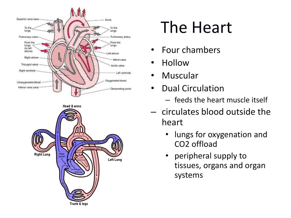
Human Anatomy and Physiology Lab (BSB 141) - Course Hero Learning Objectives. Identify all the components of the thoracic cage and be able to orient the parts correctly into their anatomical arrangements. Identify all the bones of the appendicular skeleton and know which key bone markings are unique to them. Distinguish a male pelvis from a female pelvis based on their shapes. 7.5 The Thoracic Cage - Anatomy & Physiology The thoracic cage functions to protect the heart and lungs. The sternum consists of the manubrium, body, and xiphoid process. The manubrium forms the expanded, superior end of the sternum. It has a jugular (suprasternal) notch, a pair of clavicular notches for articulation with the clavicles, and receives the costal cartilage of the first rib. The thoracic cage - the ribs and sternum | Human Anatomy and Physiology ... The thoracic cage surrounds and protects the heart and lungs in the thoracic cavity. It consists of the ribs, the sternum, and the thoracic vertebrae, to which the ribs articulate. We examined the thoracic vertebrae last lab, so here we will only examine the ribs and sternum. There are twelve pairs of ribs. Chapter 20-Cardiovascular System Flashcards - Quizlet Correctly label the following external anatomy of the anterior heart. Place the labels in order denoting the flow of blood through the pulmonary circulation beginning with the emptying of blood from the superior vena cavae. True or False: Identify which of the following are age related changes of the heart.
thoracic cavity | Description, Anatomy, & Physiology | Britannica The heart is covered by a fibrous membrane sac called the pericardium that blends with the trunks of the vessels running to and from the heart. The thoracic cavity also contains the esophagus, the channel through which food is passed from the throat to the stomach. The chest cavity is lined with a serous membrane, which exudes a thin fluid. Anatomy & Physiology: The Unity of Form and Function - Quizlet Correctly label the pathway for the cardiac conduction system. Drag each label to the location of each structure described. Vessel carrying oxygenated blood to the myocardium. Artery carrying deoxygenated blood. First vessel of systemic circuit. Veins carrying oxygenated blood. Veins carrying deoxygenated blood from the lower extremity. Correctly Label The Following External Anatomy Of The Anterior Heart In this activity, students will learn to correctly label the following external anatomy of the anterior human heart. They will understand the function of the internal and external arteries that carry blood throughout the body. The aortic sinus body and the thoracic cage also play a role in the circulatory system. Answered: 3. The heart is located within the… | bartleby Solution for 3. The heart is located within the thoracic cage, between the and rib. a. Second, fifth b. Second, third c. First, second d. First, sixth
Heart - anterior exposure | Subclavian artery, Carotid artery, Arteries ...
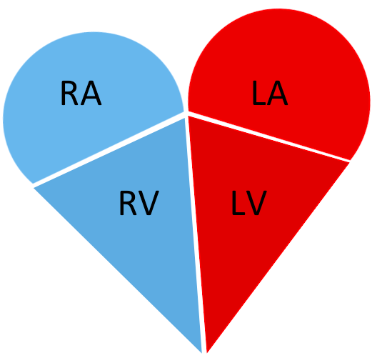
AHCDW15Notes4.pdf - Course Hero Correctly label the following anatomical features of the heart and thoracic cage. Explanation: The heart is located in the thoracic cavity in the mediastinum, between the lungs and deep to the sternum. From its superior to inferior midpoints, it is tilted toward the left, so about twothirds of the heart lies to the left of the median plane.
PPT - Leads, Leads and More Leads…. The question is.. where does this ...
(PDF) Thoracic cage - researchgate.net The purpose of the thoracic cage is to protect the organs and visceral structures within the thoracic cavity and. assist in respiration through rib movement. Mechanism of injury. Rib trauma is ...
Solved: Correctly Label The Following Anatomical Features ... | Chegg.com
The Thoracic Cage · Anatomy and Physiology The thoracic cage protects the heart and lungs. Thoracic Cage The thoracic cage is formed by the (a) sternum and (b) 12 pairs of ribs with their costal cartilages. The ribs are anchored posteriorly to the 12 thoracic vertebrae. The sternum consists of the manubrium, body, and xiphoid process.

PPT - CARDIOVASCULAR SYSTEM PowerPoint Presentation, free download - ID ...
Thorax: Anatomy, wall, cavity, organs & neurovasculature - Kenhub The thoracic, or chest wall, consists of a skeletal framework, fascia, muscles, and neurovasculature - all connected together to form a strong and protective yet flexible cage. The thorax has two major openings: the superior thoracic aperture found superiorly and the inferior thoracic aperture located inferiorly.
multiple pages cardiac cycle
Correctly Label The Following Muscles Of The Anterior View The serratus anterior muscle originates from the back and wraps around the sides of the rib cage. The serratus anterior is shown in Figure 8-8. The major muscles of the body are in the anatomical right and posterior view. The anatomical left is used to identify the superficial muscles while the anatomical right is used to label the deep ones.
Post a Comment for "41 correctly label the following anatomical features of the heart and thoracic cage."