43 label the indicated parts of the microscope
Answered: SOMATIC CELL DIVIS SPECIES, EA CH WITH… | bartleby Transcribed Image Text: SOMATIC CELL DIVISION IN 4 DI/FFERENT SPECIES, EA CH WITH A DIFFERENT NUMBER OF CIHROMOSOMES * Part IV. Images The images on the left represent events of a mitotic division in a haploid cell in which N=2. In the other boxes in each row, draw the same events for cells with the indicated chromosome number. Label-free detection and quantification of ultrafine particulate … 26.07.2022 · While it is known that air borne ultrafine particulate matter (PM) may pass through the pulmonary circulation of blood at the alveolar level between lung and heart and cross the air-blood barrier, the mechanism and effects are not completely clear. In this study the imaging method fluorescence lifetime imaging microscopy is adopted for visualization with high spatial …
Label the parts of the microscope below in the - Course Hero Label the parts of the microscope below in the corresponding numbers under the image. If you need assistance, you may refer to the labeled microscope in the following link: - microscope-parts/ Place your answers for the numbered microscope parts here: 1 Ocular is what you look through at the top of the microscope 2 Eye tube hold the eyepieces in place above the objective lense 3 Objective Lense are the primary optical lenses on a microscope 4 Nosepiece houses the objective 5 Coarse and fine ...
Label the indicated parts of the microscope
Label The Parts Of A Microscope Worksheet Answers Label the parts of the microscope indicated and state the. Can be used for practice or as a quiz. We tried to locate some good of microscope parts and use worksheet answer key along with labeling the parts of. Handphone Tablet Desktop Original Size There are many key components to understand when utilizing a microscope. Get thousands of teacher ... Parts of a microscope with functions and labeled diagram 19.04.2022 · Figure: Diagram of parts of a microscope. There are three structural parts of the microscope i.e. head, base, and arm. Head – This is also known as the body. It carries the optical parts in the upper part of the microscope. Base – It acts as microscopes support. It also carries microscopic illuminators. Visualizing Intracellular Organelle and Cytoskeletal Interactions … 15.11.2018 · Cells were imaged 16-36 hours post-transfection in a microscope stage top micro-incubator (OKO Lab) maintained at 37°C and 5% CO 2. Where indicated, the cells transfected with HaloTag plasmids were labeled with JF 646 ligand following the published protocol (Grimm et al., 2015), and the cells were imaged immediately afterward. Drosophila embryo
Label the indicated parts of the microscope. PDF Parts of the Light Microscope - Science Spot B. NOSEPIECE microscope when carried Holds the HIGH- and LOW- power objective LENSES; can be rotated to change MAGNIFICATION. Power = 10 x 4 = 40 Power = 10 x 10 = 100 Power = 10 x 40 = 400 What happens as the power of magnification increases? PDF Microscope Parts and Functions - WPMU DEV Microscope Parts and Functions Microscope One or more lenses that makes an enlarged image of an object. 8/7/2018 2 •Simple •Compound •Stereoscopic ... Always carry a microscope with one hand holding the arm and one hand under the base. Base Diaphragm A B Carrying a Microscope. 8/7/2018 13 (Solved) - Care and Structure of the Compound Microscope 1. Label all ... The Bright-field Compound ... Transport of intensity diffraction tomography with non ... - Nature 02.06.2022 · We present a new label-free three-dimensional (3D) microscopy technique, termed transport of intensity diffraction tomography with non-interferometric synthetic aperture (TIDT-NSA). Without ...
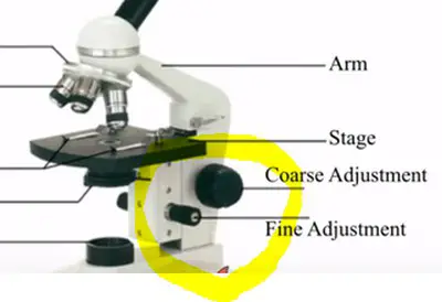
Molecular Expressions Microscopy Primer: Anatomy of the Microscope ... Microscope manufacturers offer a wide range of objective designs to meet the performance needs of specialized imaging methods, to compensate for cover glass thickness variations, and to increase the effective working distance of the objective. Learn to identify microscope objectives and their specialized properties by deciphering the engravings on the barrel. Label-free detection and quantification of ultrafine ... Jul 26, 2022 · While it is known that air borne ultrafine particulate matter (PM) may pass through the pulmonary circulation of blood at the alveolar level between lung and heart and cross the air-blood barrier, the mechanism and effects are not completely clear. In this study the imaging method fluorescence lifetime imaging microscopy is adopted for visualization with high spatial resolution and ... 6a). Identify the type of microscope shown below b). Label the parts of ... Physic question Problem 27.64 Part A A compound microscope uses a 80.0 mm lens as the objective and a 2.0 cm lens as the eyepiece. The specimen will be mounted 112 mm from the objective. Determine the barrel length. openlab.citytech.cuny.edu The Microscope EXERCSE Mar; qldb Lab Time/Date Name Care and Structure of the Compound Microscope I. Label all indicated parts of the microscope. esQi€ce 2. Explain the proper technique for transporting the microscope. 0b qs Ont en arm loose. 33 . 34 Review Sheet 3 3. Each of the following statements is either true or false.
Labeling the Parts of the Microscope | Microscope World Resources Labeling the Parts of the Microscope. This activity has been designed for use in homes and schools. Each microscope layout (both blank and the version with answers) are available as PDF downloads. You can view a more in-depth review of each part of the microscope here. Download the Label the Parts of the Microscope PDF printable version here. PDF The Microscope - Holly H. Nash-Rule, PhD Care and Structure of the Compound Microscope 1. Label all indicated parts of the microscope. Ocular lenses Rotating nosepiece Objective lenses Stage Mechanical stage Iris diaphragm lever Condenser Substage light Head Arm Power switch Light control Coarse adjustment knob Fine adjustment knob Base 2. Explain the proper technique for transporting the microscope. Lungs (Human Anatomy): Picture, Function, Definition, Conditions - WebMD WebMD's Lungs Anatomy Page provides a detailed image and definition of the lungs. Learn about lung function, problems, location in the body, and more. 07-894-5553 | 15 g, 12/Pkg | Neo-Predef® with Tetracaine Powder 12.02.2015 · Neo-Predef® with Tetracaine Powder, 15 g, 12/Pkg, ZOETIS, Manufacturer Item #:10000815, Patterson Item #:07-894-5553
Untitled
Transport of intensity diffraction tomography with non ... Jun 02, 2022 · Since its invention in the 1600s, the optical microscope has experienced continuous development and become an indispensable tool for the visualization of micro-scale objects with high resolution ...
microscope | Types, Parts, History, Diagram, & Facts | Britannica
Solved > 1. Label all indicated parts of the microscope. from Chapter 3 ... Solved expert answers for Human Anatomy and Physiology Laboratory Manual, Main Version 10th Edition by Elaine N. Marieb, Susan J. Mitchell, Lori A. Smith. Instant access with 24/7 expert assistance.

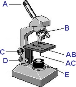
This is a common compound microscope. Label its parts from A ...
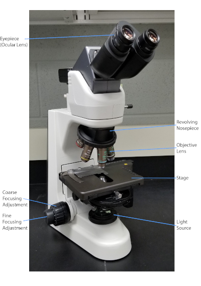
Microscope Parts & Functions - AmScope Nosepiece: The upper part of a compound microscope that holds the objective lens. Also called a revolving nosepiece or turret. Also called a revolving nosepiece or turret. Numerical Aperture (N.A): A measure of the diameter of the aperture compared to the focal length of a lens and ultimately, of the resolving power of a microscope.

Compound Microscope: Parts of Compound Microscope
Virtual Microscope - NCBioNetwork.org Lesson Description BioNetwork’s Virtual Microscope is the first fully interactive 3D scope - it’s a great practice tool to prepare you for working in a science lab. Explore topics on usage, care, terminology and then interact with a fully functional, virtual microscope. When you are ready, challenge your knowledge in the testing section to see what you have learned.
Parts of the Microscope Quiz
Microscope Quiz: How Much You Know About Microscope Parts ... - ProProfs Projects light upwards through the diaphragm, the specimen, and the lenses. 5. Is used to regulates the amount of light on the specimen. Supports the slide being viewed. Moves the stage up and down for focusing. 6. Is used to support the microscope when carried. Moves the stage slightly to sharpen the image.

PPT - Label the parts on your microscope picture. PowerPoint ...
UD Virtual Compound Microscope - University of Delaware ©University of Delaware. This work is licensed under a Creative Commons Attribution-NonCommercial-NoDerivs 2.5 License.Creative Commons Attribution-NonCommercial-NoDerivs 2.5 …

An Introduction to the Light Microscope, Light Microscopy ...
A. Label the parts of the microscope below. Identify whether the ... 3. Illuminator - Illuminator is the light source for a microscope. Mechanical: 1. Base - It helps in holding the various parts of microscope. It also contains a light source. 2. Arm - It is used for holding the microscope. And which is connected the eyepiece to the objective lens. 3. Stage - It is a rigid platform on which specimen to be viewed ...
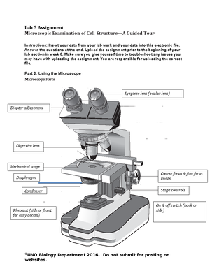
Lab 5 Assignment - Lab 5 Assignment Microscopic Examination ...
Virtual Microscope - NCBioNetwork.org Lesson Description BioNetwork’s Virtual Microscope is the first fully interactive 3D scope - it’s a great practice tool to prepare you for working in a science lab. Explore topics on usage, care, terminology and then interact with a fully functional, virtual microscope.
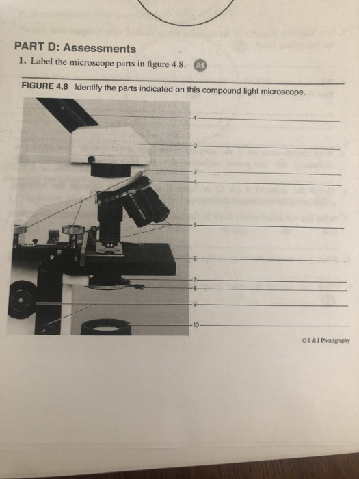
Solved PART D: Assessments 1. Label the microscope parts in ...
Label parts on microscope Flashcards | Quizlet Terms in this set (14) Eyepiece. Body Tube on microscope. High power objective. Low power objective. Stage Opening. Diaphragm Lever. Light on microscope. Coarse adjustment.

Tracking the formation and degradation of fatty-acid ...
SOLVED:Use the information below to label the parts of the microscope ... Let's get started. A So, for a we have the outside of the eye of a white region. This is Venus Clara. Well, B next, we have the the back of the eye for region that detects light. This is for retina. See, this is pointing to the vessels and coming into the eye, but provides blood. So is so is D. And if you look very closely see is pointing for ...
BIO 101 parts of the microscope to label - NAME Click or tap ...
Educational Atomic Force Microscope (AFM) - Thorlabs Nov 05, 2021 · Thorlabs' Atomic Force Microscope Kits are available in imperial and metric versions. In cases where the metric and imperial kits contain parts with different item numbers, metric part numbers and measurements are indicated by parentheses unless otherwise noted.

ZEISS Axio Observer for Life Science Research
Educational Atomic Force Microscope (AFM) - Thorlabs 05.11.2021 · Thorlabs' Atomic Force Microscope Kits are available in imperial and metric versions. In cases where the metric and imperial kits contain parts with different item numbers, metric part numbers and measurements are indicated by parentheses unless otherwise noted.
Parts of a Microscope Quiz
LABEL ALL INDICATED PARTS OF THE MICROSCOPE.docx - Course Hero LABEL ALL INDICATED PARTS OF THE MICROSCOPE. 2. LABEL ALL INDICATED PARTS OF THE MICROSCOPE. 3. LABEL ALL INDICATED PARTS OF THE MICROSCOPE. 4 LABEL ALL INDICATED PARTS OF THE MICROSCOPE. 5 LABEL ALL INDICATED PARTS OF THE MICROSCOPE. 6 EXPLAIN THE PROPER TECHNIQUE FOR TRANSPORTING THE MICROSCOPE. WHEN TRANSPORTING THE MICROSCOPE, HOLD IT IN AN UPRIGHT POSITION WITH ONE HAND ON ITS ARM AND THE OTHER SUPPORTING ITS BASE.
Parts of the Microscope with Labeling (also Free Printouts ...
The Lungs (Human Anatomy): Picture, Function, Definition ... Sputum cytology: Viewing sputum under a microscope for abnormal cells can help diagnose lung cancer and other conditions. Lung biopsy: A small piece of tissue is taken from the lungs, either ...
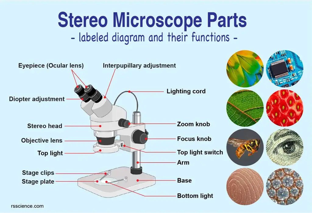
Parts of Stereo Microscope (Dissecting microscope) – labeled ...
Parts of the Microscope with Labeling (also Free Printouts) Parts of the Microscope with Labeling (also Free Printouts) By Editorial Team March 7, 2022 A microscope is one of the invaluable tools in the laboratory setting. It is used to observe things that cannot be seen by the naked eye. Table of Contents 1. Eyepiece 2. Body tube/Head 3. Turret/Nose piece 4. Objective lenses 5. Knobs (fine and coarse) 6.

Compound Microscope Parts
Solved Care and Structure of the Compound Microscope 1. - Chegg Label all indicated parts of the microscope. Cvlar len hy 3. The following statements are true or false. If true, write ing on the blank the proper word or phrase to replace the one that is underlined. Ton the answer blank. If false, correct the statement by writ 1. The microscope lens may be cleaned with any soft tissus 2.

Microscope Labeling Diagram | Quizlet
PDF Parts of the compound microscope: Write the correct label for each part ... Parts of the compound microscope: Write the correct label for each part of the microscope shown below: Using the compound microscope: Match each part of the compound microscope on the left with its function on the right. 13. base and arm A. eyepiece, what you look in to see an image
Microscopy and Staining Techniques in Bacteria | Microbiology ...
Solved Care and Structure of the Compound Microscope 1. - Chegg Biology questions and answers. Care and Structure of the Compound Microscope 1. Label all indicated parts of the microscope. H B D K L F < G N 2. Explain the proper technique for transporting the microscope. Question: Care and Structure of the Compound Microscope 1. Label all indicated parts of the microscope. H B D K L F < G N 2.
Special Sense organ
Microscope Parts and Functions Arm: The arm connects the body tube to the base of the microscope. Coarse adjustment: Brings the specimen into general focus. Fine adjustment: Fine tunes the focus and increases the detail of the specimen. Nosepiece: A rotating turret that houses the objective lenses.
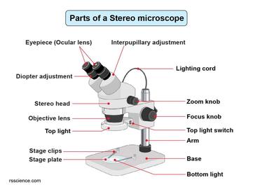
Parts of Stereo Microscope (Dissecting microscope) – labeled ...
UD Virtual Compound Microscope - University of Delaware ©University of Delaware. This work is licensed under a Creative Commons Attribution-NonCommercial-NoDerivs 2.5 License.Creative Commons Attribution-NonCommercial-NoDerivs 2

microscope | Types, Parts, History, Diagram, & Facts | Britannica
Compound Microscope: Parts of Compound Microscope - BYJUS The parts of the compound microscope can be categorized into: Mechanical parts; Optical parts (A) Mechanical Parts of a Compound Microscope. 1. Foot or base. It is a U-shaped structure and supports the entire weight of the compound microscope. 2. Pillar. It is a vertical projection. This stands by resting on the base and supports the stage. 3. Arm
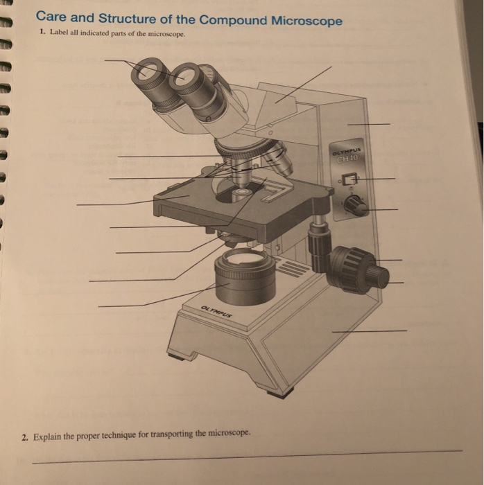
Solved Care and Structure of the Compound Microscope 1 ...
Quia - Label the Microscope Quiz Label the Microscope Quiz. Choose the word that correctly labels the parts of the microscope. Please enter your name. First name: Last name . Tools. Copy this to my account; E-mail to a friend; Find other activities; Start over; Print; Help; Mrs. Coyle. Simmons Elementary School. Versailles, KY:
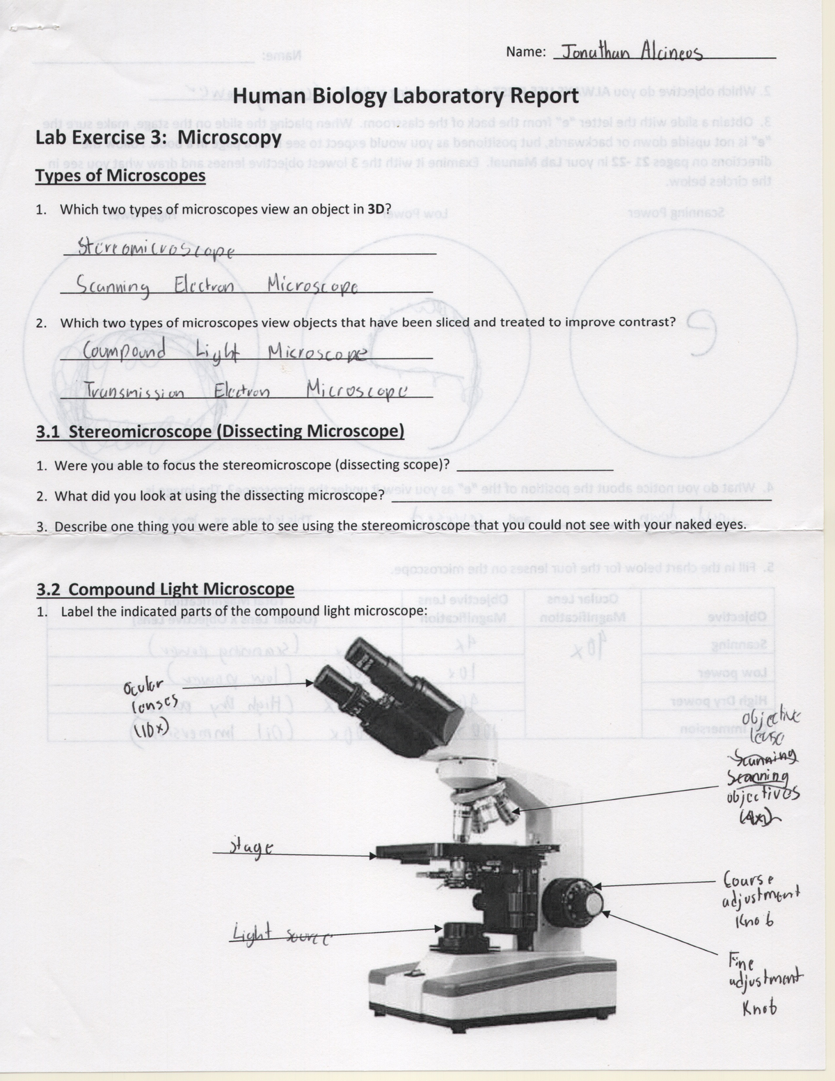
Lab Exercise 3 Human Biology Lab Report - BSC 1020 - StuDocu
The Microscope - City University of New York Name Care and Structure of the Compound Microscope I. label alt indicated parts of the microscope oos¿piece. 2. the proper techntque for transporting the microscope one hand arm . 33. Review Sheet 3 34 3. Each of the following statements IS either true or false If true, write T on the answer blank If false, correct the statement by writing on the blank the proper word or phrase to replace the one that IS underlined 02 3 4 The microscope lens may be cleaned The microscope should be stored ...
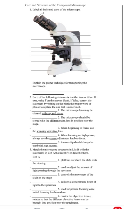
Solved Care and Structure of the Compound Microscope 1 ...
Label the microscope — Science Learning Hub In this interactive, you can label the different parts of a microscope. Use this with the ...
INSIRUCIIONS
MasteringMicrobiology - Week 1 Post Lab Flashcards | Quizlet Label parts of the microscope. (From top to bottom) Ocular Objective Stage Condenser Lamp Label parts of the microscope. (From top to bottom) Objective Lens Slide Clip Stage Label the indicated parts of the microscope. (From top to bottom and left to right) Ocular Lens Objective Lens Oil Immersion Lens Navigation Knobs Coarse Focus Fine Focus

MICRO - Georgia State University - Course Hero
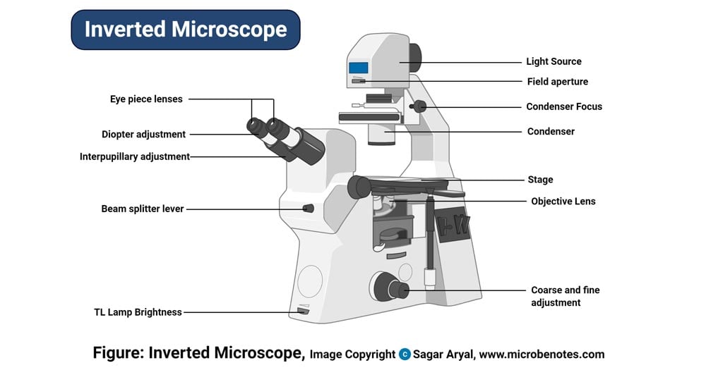
Parts of a microscope with functions and labeled diagram - Microbe Notes Apr 19, 2022 · Figure: Diagram of parts of a microscope. There are three structural parts of the microscope i.e. head, base, and arm. Head – This is also known as the body. It carries the optical parts in the upper part of the microscope. Base – It acts as microscopes support. It also carries microscopic illuminators.

This is a common compound microscope. Label its parts from A ...
Visualizing Intracellular Organelle and Cytoskeletal Interactions … 15.11.2018 · Cells were imaged 16-36 hours post-transfection in a microscope stage top micro-incubator (OKO Lab) maintained at 37°C and 5% CO 2. Where indicated, the cells transfected with HaloTag plasmids were labeled with JF 646 ligand following the published protocol (Grimm et al., 2015), and the cells were imaged immediately afterward. Drosophila embryo

This is a common compound microscope. What the labelling D ...
Parts of a microscope with functions and labeled diagram 19.04.2022 · Figure: Diagram of parts of a microscope. There are three structural parts of the microscope i.e. head, base, and arm. Head – This is also known as the body. It carries the optical parts in the upper part of the microscope. Base – It acts as microscopes support. It also carries microscopic illuminators.
Parts of a microscope with functions and labeled diagram
Label The Parts Of A Microscope Worksheet Answers Label the parts of the microscope indicated and state the. Can be used for practice or as a quiz. We tried to locate some good of microscope parts and use worksheet answer key along with labeling the parts of. Handphone Tablet Desktop Original Size There are many key components to understand when utilizing a microscope. Get thousands of teacher ...

Basic Geology 1. Crystallography. - ppt download

Semi automatic Manual Marking Machine YL 360 Sign Nameplate ...

Compound Microscope Parts – Labeled Diagram and their ...

Labeling the Parts of the Microscope | Microscope activity ...
Solved] Pleas answer all and thanks | Course Hero

Cool 30 Top For Simple Microscope Diagram References - Matahari
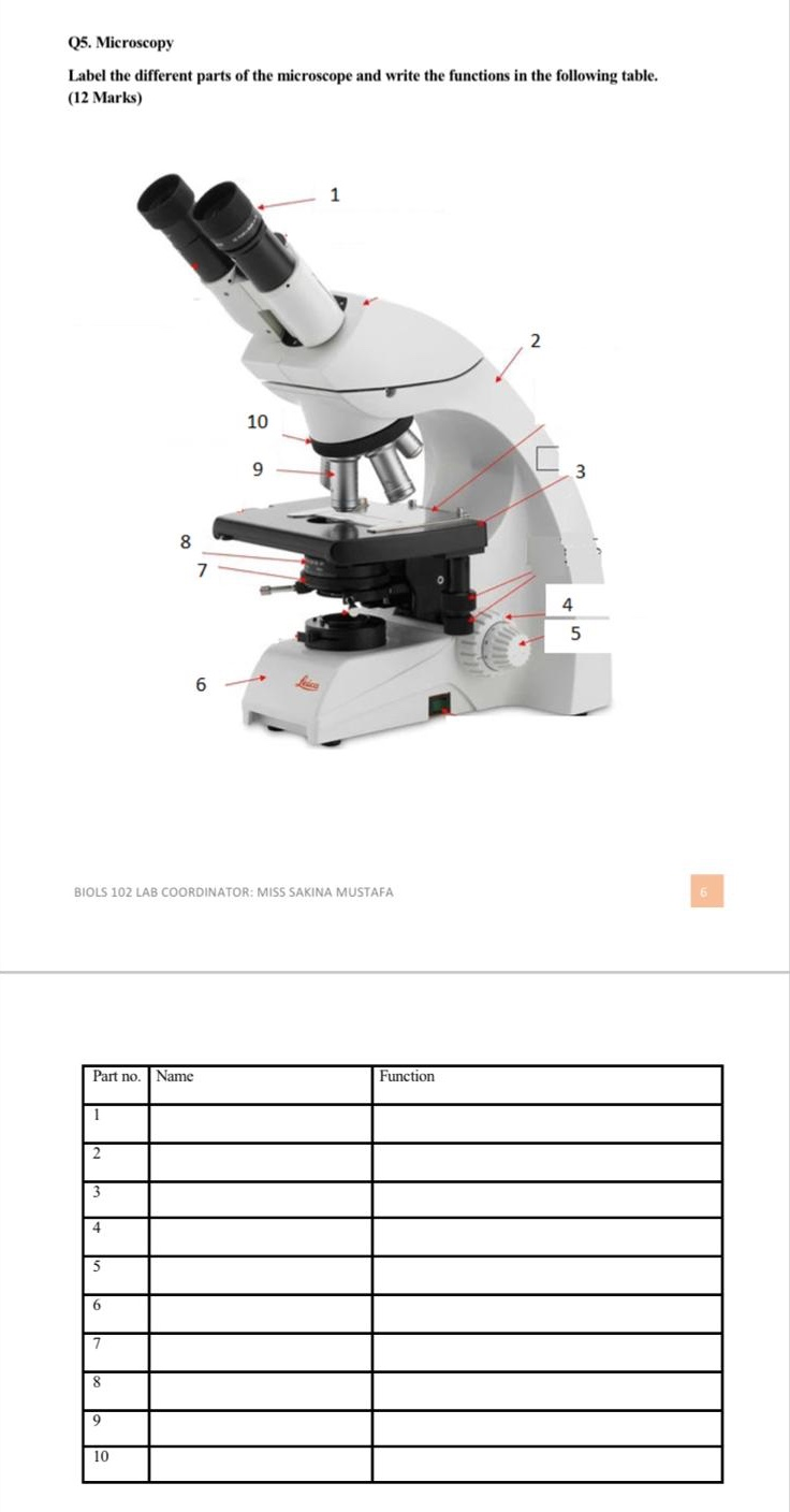
Solved Q5. Microscopy Label the different parts of the ...
Microscope Quiz

Compound Microscope Parts – Labeled Diagram and their ...
Brainstem: Definition, anatomy, parts, function | Kenhub

Microscope Diagram Labeled, Unlabeled and Blank | Parts of a ...

Care and Structure of the Compound Microscope 1.jpg - Care ...

Follow J*'s (@Afri_Abana) latest Tweets / Twitter
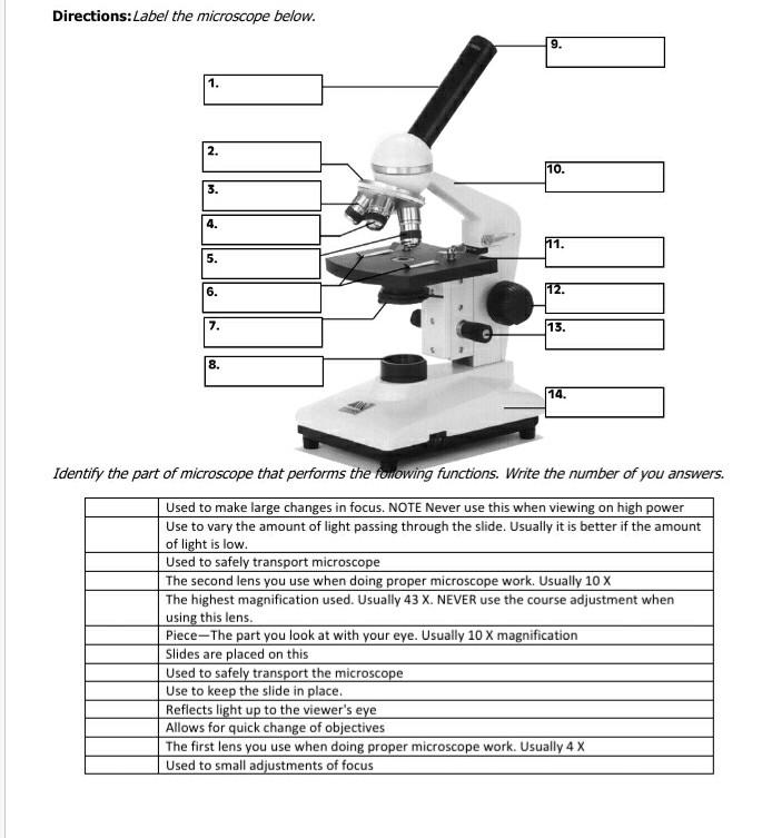
Solved Directions:Label the microscope below. 9. 1. 111 2 ...
Post a Comment for "43 label the indicated parts of the microscope"