44 cow eye dissection labeled
anatomy of the eye labeled Cow Eye Dissection - YouTube we have 7 Pictures about Cow Eye Dissection - YouTube like Image result for Eye Model Labeled | Ear anatomy, Eyes, Ocular, Cow Eye Dissection - YouTube and also Untitled Document [bio.sunyorange.edu]. Here you go: Cow Eye Dissection - YouTube cow eye dissection Cow's Eye Dissection - Eye diagram - Exploratorium A muscle that controls how much light enters the eye. It is suspended between the cornea and the lens. A cow's iris is brown. Human irises come in many colors, including brown, blue, green, and gray. A clear fluid that helps the cornea keep its rounded shape. The pupil is the dark circle in the center of your iris.
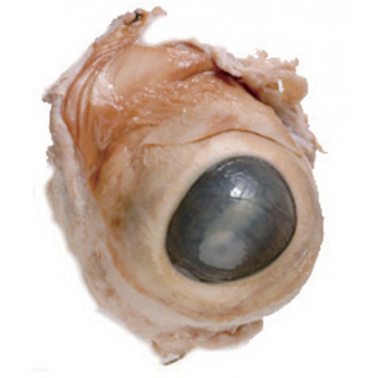
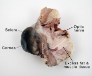
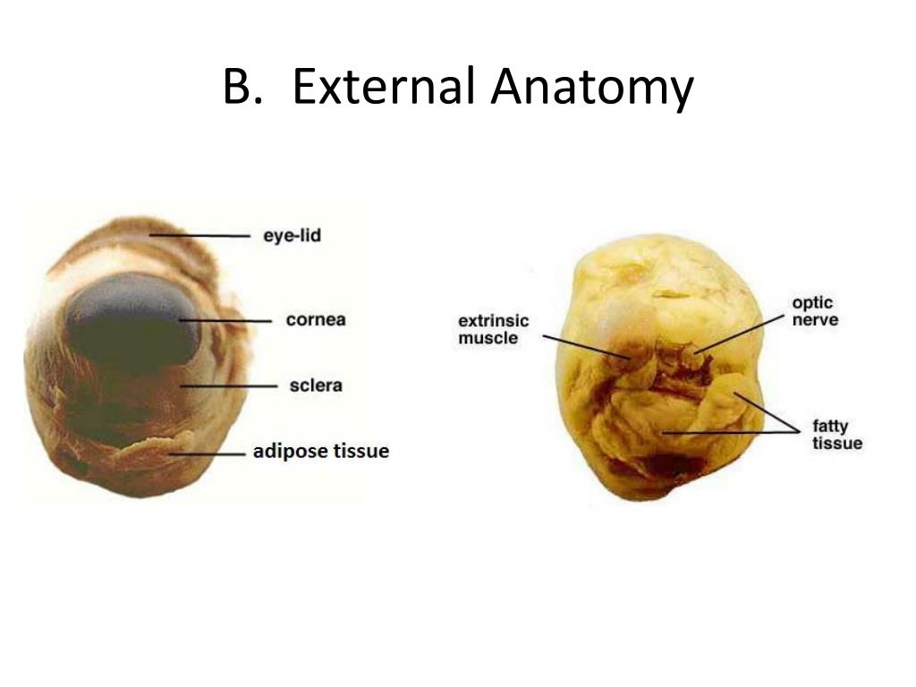
Cow Eye Dissection Lab from Anatomy and Physiology Safety goggles • Dissection scissors. Preserved cow's eye • Dissection tray. Forceps • Vinyl or latex gloves. Dissection probes. Apron or tee-shirt; Paper towels. Plastic trash bag. External features of the eye. A. Locate the cornea, sclera, and optic nerve. a. The white part of the eye, the sclera, is a tough, outer covering of the ...
Cow eye dissection labeled
PDF Cow Eye Dissection Guide - Central Bucks School District DISSECTION OF THE COW EYE Please make sure to wear gloves and safety glasses when you are dissecting, and make sure to clean up thoroughly after the lab. Also, the cow eyes can be rather slippery, so use caution when handling and cutting them. You will need a scalpel and forceps. 1. First, identify the most external structures of the eye. Cow Eye Dissection - YouTube About Press Copyright Contact us Creators Advertise Developers Terms Privacy Policy & Safety How YouTube works Test new features Press Copyright Contact us Creators ... anatomy of a cow eye Cow Eye Dissection Labeled | Cow Eye Sagittal Section | Biology 100 . eye dissection cow human anatomy retina body eyes science choroid labeled coat guide hometrainingtools project physiology learn biology blind spot. Anatomy, physiology, and pathophysiology. Dissection labeled dissecting labo.
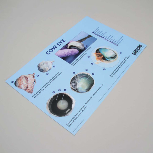
Cow eye dissection labeled. Cow Eye Dissection Teaching Resources | Teachers Pay Teachers This is a comprehensive dissection guide of the cow eye, designed for a high school or early college Biology or Anatomy & Physiology class. The guide includes step-by-step instructions and labeled diagrams that will lead students through the external anatomy of the eye, followed by dissection of the internal structures. cow internal anatomy Cow's eye dissection. Eye diagram cow eyes anatomy labeled exploratorium human dissection edu chart learning cliparts biology physiology med museum science muscles animal. Planarian system digestive platyhelminthes hydra structure internal phylum cavity gastrovascular simple freshwater felina mouth cnidaria Cow eye - dissection and label Cow eye shown with labeled cornea. The cornea is the transparent front part of the eye that covers the iris, pupil, and anterior chamber. The cornea, with the anterior chamber and lens, refracts light, with the cornea accounting for approximately two-thirds of the eye's total optical power. 3. Cow Eye Dissection & Anatomy Project | HST Learning Center Cow Eye Dissection: Internal Anatomy 1. Place the cow's eye on a dissecting tray. The eye most likely has a thick covering of fat and muscle tissue. Carefully cut away the fat and the muscle. As you get closer to the actual eyeball, you may notice muscles that are attached directly to the sclera and along the optic nerve.
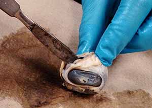
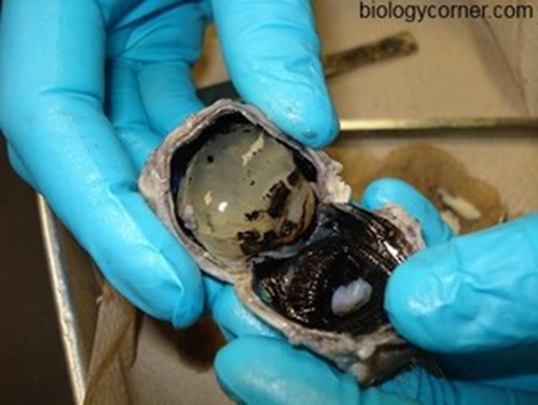
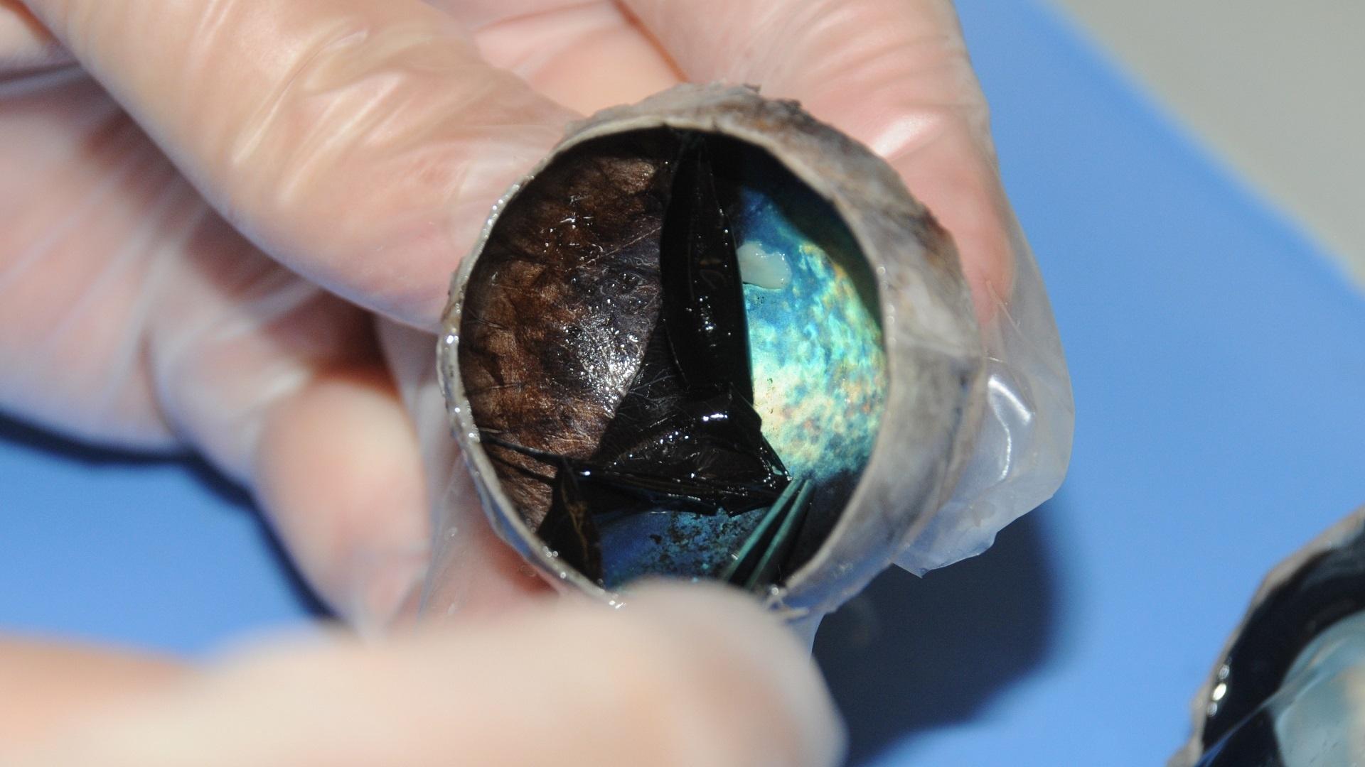
anatomy of a cow eye Fresh cow eye dissection (Part 1): Preparation - YouTube we have 9 Pics about Fresh cow eye dissection (Part 1): Preparation - YouTube like Prof. Wilson sheep eye dissection. ... Brain sheep dissection anatomy structures labeled cerebral human fissure transverse physiology biology cerebellum cerebrum diagram sulci lobe gyri cortex mater. Sheep ... Cow Eye Dissection | Carolina.com Cow Eye Internal Anatomy Hold the eye between your thumb and forefinger, as shown below. Using scissors or a scalpel, carefully cut the eye in half, separating the front and back of the eye. Examine the inside front portion of the eye. Remove the gelatinous vitreous humor and hard lens. Dissection Lab Report - APHY101 - Ivy Tech - StuDocu Abnormal Psychology (PSY 411) Communication at Work (COM 100) Anatomy and Physiology 1 (APHY-101) Macroeconomics (ECON0110) Criminal Justice 230 (CJ230) Survey of Asian Empires (HIS-350) Forensic Science (BLAW2) Methods and Strategies for Teaching English Language Arts (elm-480) Documents. Cow Eye Dissection Guide - Google Slides Cow Eye. Use the point of a scissors or a scalpel to make an incision through the layers of the eye capsule (similar to figure 1); there are three layers from the exterior: sclera, whitish/grey, continuous with the transparent cornea, choroid, thin dark black layer and the retina, thin greyish/pink layer. Use a scissors to dissect the entire ...
cow brain anatomy cow eye dissection labeled parts anatomy eyes labels label section diagram structures virtual physiology lab human resize science body tapetum Brain Of A Cow Stock Photo. Image Of Activity, Abstract - 45875504 brain cow background A Day In The Life Of A Cow Vet: She's Crazy!!! cowdoc1981.blogspot.com PDF Cow Eye Dissection: Examining Structure and Function The eyes of cows are structurally and functionally similar to the eyes of humans. During this activity, you will dissect a cow eye. You will observe several important features of the eye and develop your understanding of how each part functions to make vision possible. Materials Cow Eye Dissection - The Biology Corner The cow eye is a fantastic specimen for students of all ages to dissect. The structures are clear, dissection easy to accomplish and usually kids enjoy the lab. The lab guide for students outlines the procedure for the dissection and you can view the eye gallery to see photographs of the dissection. Cow Eye Dissection Kit for Kids Animal Anatomy Labs | HST Inside this cow eye dissection kit, you'll find: a preserved cow eye, a full-color photographic eye dissection guide, and the dissection tools/lab equipment you'll need: a #22 broad-blade scalpel, scissors, and a sturdy disposable dissecting tray. You'll get see how a cow's eye is like a human eye as you closely investigate parts from the iris ...
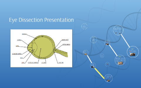
Eye Dissection Presentation by Joshua Lee
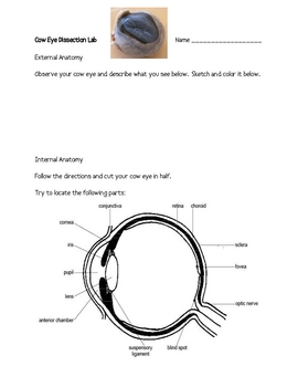
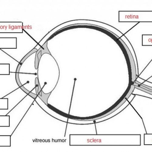
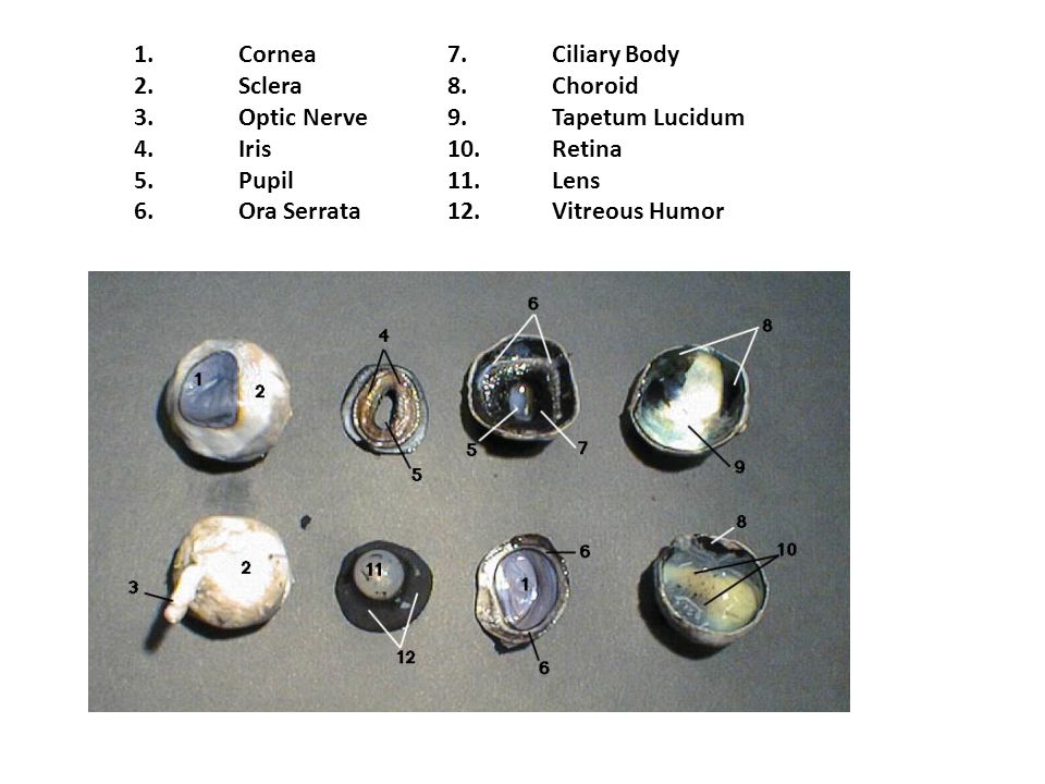
PDF Name: Dissection 101: Cow Eye human eye _____ _____ Draw and label the cow eye. Cornea Optic nerve Vitreous humor Retina Optic disc (blind spot) Choroid Tapetum lucidum Sclera Aqueous humor Suspensory ligaments Lens Ciliary body Pupil Iris Provided by Dissection 101: Cow Eye

Cow Eye Dissection Vocabulary - K6 Flashcards | Quizlet
Lab: Cow Eye Dissection Flashcards | Quizlet Lab: Cow Eye Dissection STUDY Flashcards Learn Write Spell Test PLAY Match Gravity Created by Daniel_Kinsey Terms in this set (14) Anterior Chamber Ciliary Muscle enabling changes in lens shape for light focusing Retina convert the light into neural signals, and send these signals on to the brain for visual recognition Choroid

Cow Eye Dissection by tiggerbaby1122 on DeviantArt
PDF Cow Eye Dissection Lab - Home Science Tools This cow eye dissection kit comes with everything you need to conduct a lab examination. Safety Guidelines • Work in a place separate from eating and food preparation areas. • Use disposable latex gloves or nitrile gloves during the dissection and cleanup. • Use only dissection tools provided.

Mrs. Knudson did her famous cow eye... - Vernon Middle School ...
Cow Eye Dissection Quiz - PurposeGames.com 0:00.0. For You. Badges (66) Tournaments (37) AI Stream The more you play, the more accurate suggestions for you. Cities by Landmarks 11p Image Quiz. Cities of Midwestern US 32p Image Quiz. I spy on... 26p Image Quiz. The Western States 11p Image Quiz.
Cow Eyes, Preserved, Pkg. of 5 | Flinn Scientific
Lab 10—Labeled Cow Eye Lab 10—Labeled Cow Eye BIO 137 Virtual Lab 10 The Cow Eye (Labeled) Return to: Lab 10 PageBIO 137 Main Page Be sure to practice using the unlabeled images. Coronal section, anterior view (lens and vitreous humor displaced) Sagittal section This page created and maintained by Udo M. Savalli. Last updated April 18, 2006.
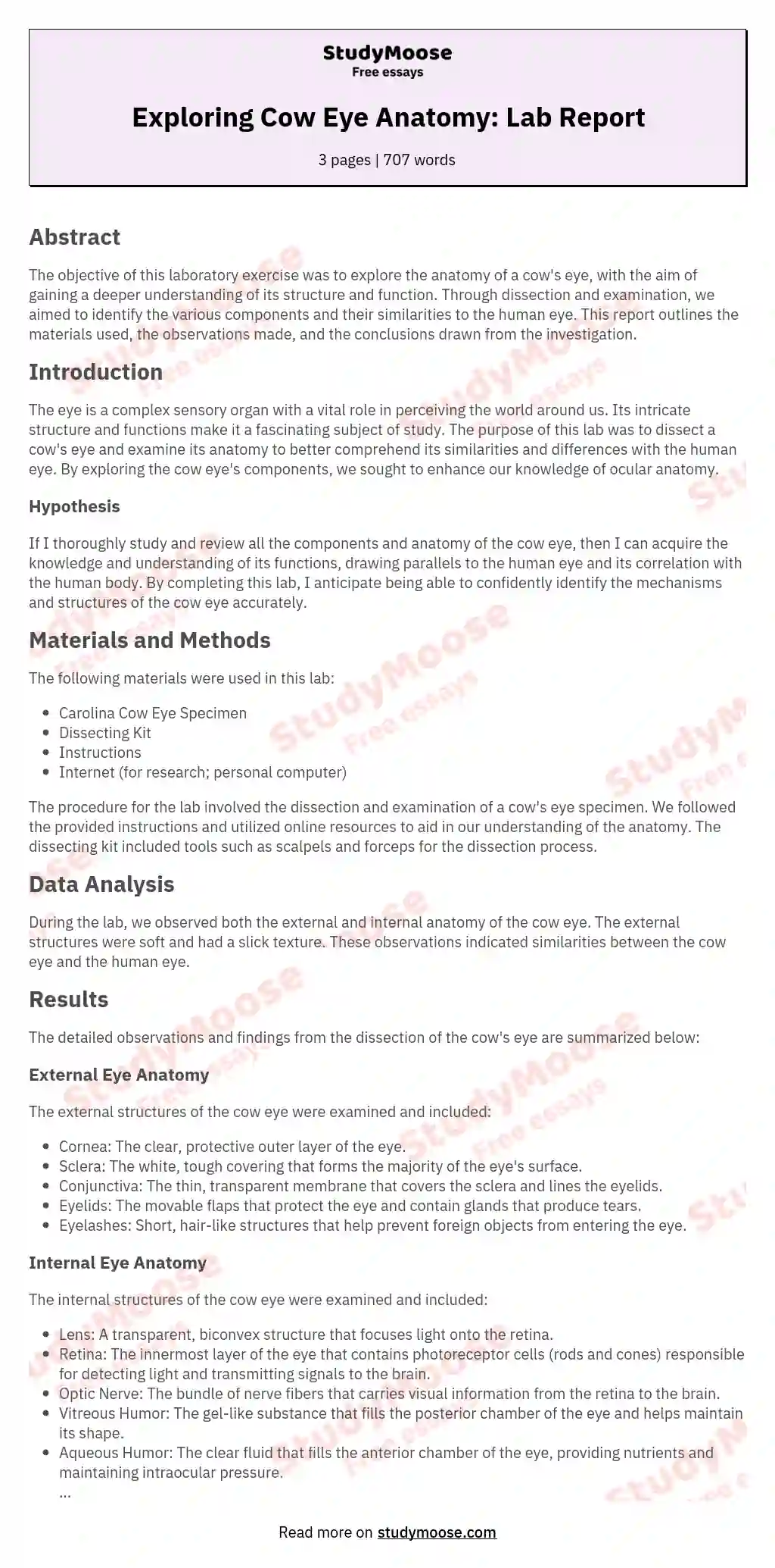
Cow Eye dissection Free Essay Example
Cow Eye Dissection & Parts of the Eye Diagram | Quizlet iris A ring of muscle tissue that forms the colored portion of the eye; controls the size of the pupil opening. retina Located in the back of the eye, contains the rods and cones. pupil The opening in the center of the iris through which light enters the eye lens
Cow Eye Dissection Lab for Elementary
sheep eye anatomy eye anatomy cow human dissection lab nervous structure labeled diagram parts anterior tunic spinal cord system internal external biology physical. Smith, k / anatomy and physiology activities. Heart right atrium ventricle anatomy labeled side internal interior chordae open tendineae structures valve tricuspid chamber chambers muscles papillary ...
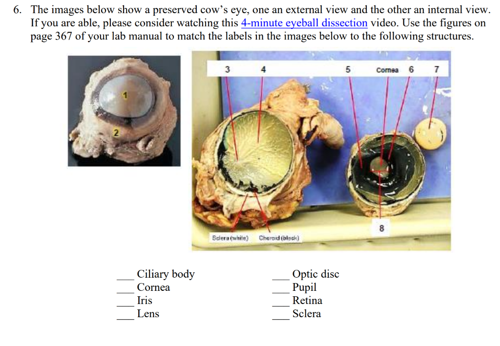
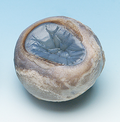
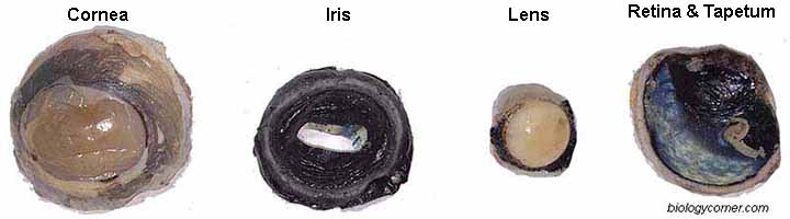
Solved 6. The images below show a preserved cow's eye, one ...
Cow Eye Dissection - The Biology Corner COW EYE DISSECTION 1. Examine the outside of the eye. You should be able to find the sclera, or the whites of the eye. This tough, outer covering of the eyeball has fat and muscle attached to it 2. Locate the covering over the front of the eye, the cornea. When the cow was alive, the cornea was clear.

Cow Eye Dissection | Carolina.com
PDF COW'S EYE dissection - Exploratorium COW'S EYE dissection page 6 Now take a look at the rest of the eye. If the vitreous humor is still in the eyeball, empty it out. On the inside of the back half of the eyeball, you can see some blood vessels that are part of a thin fleshy film. That film is the retina. Before you cut the eye open, the vitreous humor
Cow Eye Dissection: Examining Structure and Function
[Solved] How to label a cow eye dissection? | Course Hero When labeling a cow eye dissection, it is also important to use clear and concise labels. Use abbreviations if necessary, but make sure that the labels are still understandable. For example, you might label the cornea as "C" and the iris as "I". In general, it is best to label the parts of the eye before you start the dissection.
NEUR 320: Art and Vision
anatomy of a cow eye Cow Eye Dissection Labeled | Cow Eye Sagittal Section | Biology 100 . eye dissection cow human anatomy retina body eyes science choroid labeled coat guide hometrainingtools project physiology learn biology blind spot. Anatomy, physiology, and pathophysiology. Dissection labeled dissecting labo.
Cow Eye, Preserved, Pail of 50
Cow Eye Dissection - YouTube About Press Copyright Contact us Creators Advertise Developers Terms Privacy Policy & Safety How YouTube works Test new features Press Copyright Contact us Creators ...
Cow Eye Dissection & Anatomy Project | HST Learning Center
PDF Cow Eye Dissection Guide - Central Bucks School District DISSECTION OF THE COW EYE Please make sure to wear gloves and safety glasses when you are dissecting, and make sure to clean up thoroughly after the lab. Also, the cow eyes can be rather slippery, so use caution when handling and cutting them. You will need a scalpel and forceps. 1. First, identify the most external structures of the eye.
Cow Eye Dissection
Cow eye dissection mat 28cm x 43cm
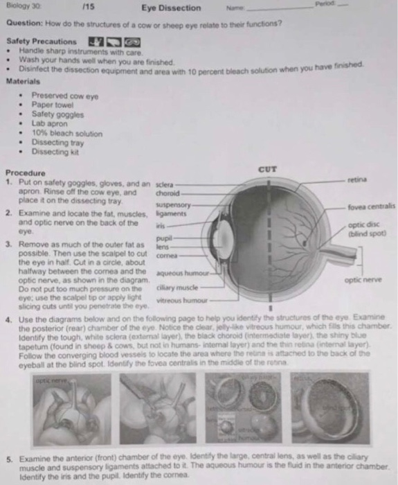
Solved retina Biology 30 15 Eye Dissection Name Question ...

Cow Eye Dissection 2 Diagram | Quizlet
Cow Eye Dissection

Eye Dissection

Cows Eyeball | PDF | Human Eye | Eye

Berkas:Schematic diagram of the human eye en.svg - Wikipedia ...

HBS 2.4.1: Anatomy of the Eye Quiz - Quizizz
![Eye Dissection || The Eyes Have It [EDU]](https://i.ytimg.com/vi/uJXkrdA87XQ/maxresdefault.jpg)
Eye Dissection || The Eyes Have It [EDU]

Cow's Eye Dissection | Exploratorium Video
Sheep Eye Dissection
Cow Eye Dissection Lab

Cow Eye Dissection Diagram | Quizlet

Diseksi Anatomi Sapi Mata manusia, luminescent blue glow iris ...
PPT - Sheep Eye Dissection PowerPoint Presentation, free ...
Cow Eye Dissection: Examining Structure and Function
Cow Eye Labeled - ClipArt Best - ClipArt Best - ClipArt Best

Cow Eye Dissection Guide - Google Slides

Classical Conversations Cow Eye Dissection - Ben and Me

Transverse section of the human eye (Wikimedia Commons, 2007 ...
SCB209 - Lab3 - Natural Sciences Open Educational Resources

Cow's Eye Dissection - Eye diagram
13.7: Cow Eye Dissection - Biology LibreTexts

Cow Eye Dissection | Carolina.com

Diagram of human eye anatomy with label 1868583 Vector Art at ...

Carolinas Perfect Solution Cow Eyes

Cow Eye Dissection | Carolina.com
Dissection 101 | Detailed Cow Eye Dissection Video (Part 1 of 2)
Sheep Eye Dissection. - ppt video online download

Cow Eye Dissection Kit
Post a Comment for "44 cow eye dissection labeled"