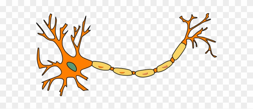
38 neuron labels
What Is a Neuron? Diagrams, Types, Function, and More - Healthline Neurons, also known as nerve cells, send and receive signals from your brain. While neurons have a lot in common with other types of cells, they're structurally and functionally unique. Specialized... Pics Of Labeled Of A Neuron Pictures, Images and Stock Photos Medical labeled diagram with all kind cells. Example of blood, neurons, cardiac, bone, intestinal, epithelial, fat, liver and muscle. The nervous system The human nervous system vector medical illustration Labeled diagram of the neuron Labeled diagram of the neuron, nerve cell that is the main part of the nervous system.
Neuron Labeling Quiz - PurposeGames.com This is an online quiz called Neuron Labeling. There is a printable worksheet available for download here so you can take the quiz with pen and paper. Your Skills & Rank. Total Points. 0. Get started! ... Label the common combining forms for the eye: 7p Image Quiz. Body Regions Labeling 33p Image Quiz. Alimentary Canal 16p Image Quiz. Accessory ...

Neuron labels
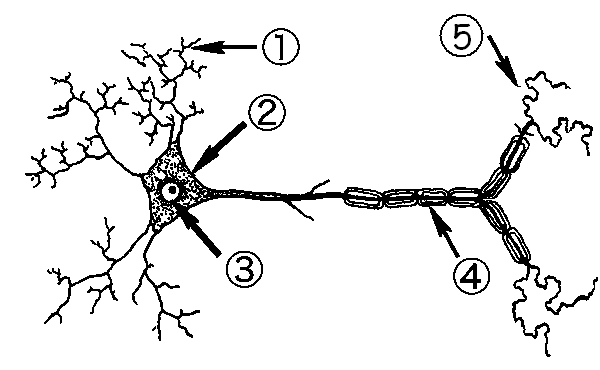
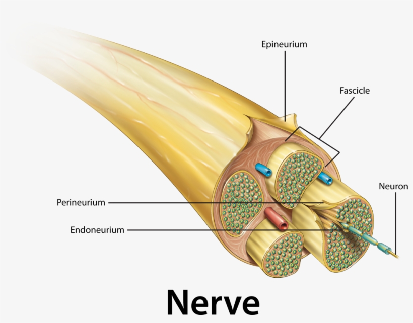
Diagram Quiz on Neuron Structure and Function (Labeling Quiz) Diagram Quiz on DNA replication. 1. Identify the cell type in the above figure. 2. In the figure, labeled '1' receives impulses from adjacent neuron. It is called the. 3. In the figure, labeled '2' is the short filaments from the cell body that carries impulses from dendrites to the cell body which is the. 4. Neuron Markers - BioLegend Neurons have highly compartmentalized structures that are generally classified into soma (cell body), axon, dendrite, axonal terminal, and synapse. BioLegend offers antibodies against markers that are expressed in each structural unit of a neuron, and allow their identification using applications such as microscopy, including immunohistochemistry (IHC) or immunocytochemistry (ICC). A Labelled Diagram Of Neuron with Detailed Explanations - BYJUS They are found in the brain, spinal cord and the peripheral nerves. A neuron is also known as the nerve cell. The structure of a neuron varies with their shape and size and it mainly depends upon their functions and their location. Neurons are the structural and functional units of the nervous system. A group of neurons forms a nerve.
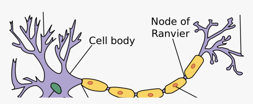
Neuron labels. Neuron Label Worksheet Teaching Resources | Teachers Pay Teachers it can be used with any biology, anatomy & physiology, psychology, or ap psychology course.the worksheet contains six major sections: 1) students color and label a neuron, 2) students complete a diagram of the subdivisions of the nervous system and answer some function questions, 3) students complete a neurotransmitter chart, 4) students make … Neuron Label And Color Teaching Resources | Teachers Pay Teachers This is a 15 page PowerPoint slideshow on the basics of a neuron. It encourages the student to create a diagram, label, and color it. Then 5-6 links are provided where the student can practice on various neuron labeling websites. At the end is an edible and play doh model of the neuron to encourage further projects. Subjects: General Science Label Parts of a Neuron Diagram | Quizlet The chemical signals that neurons use to communicate with other neurons and cells. It is the chemical involved in impulse transmission Neuron transportation Neurons generally transport signals in one direction from the dendrites, through the soma, along the axon and unto the terminal buttons Nodes of Ranvier Neurons - Neuron (labels) - histology slide Neuron (labels) - histology slide This is a histology slide of a neuron. Histology slide courtesy of William L. Todt, Ph.D. at Concordia College, Moorhead, Minnesota.
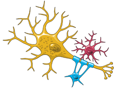
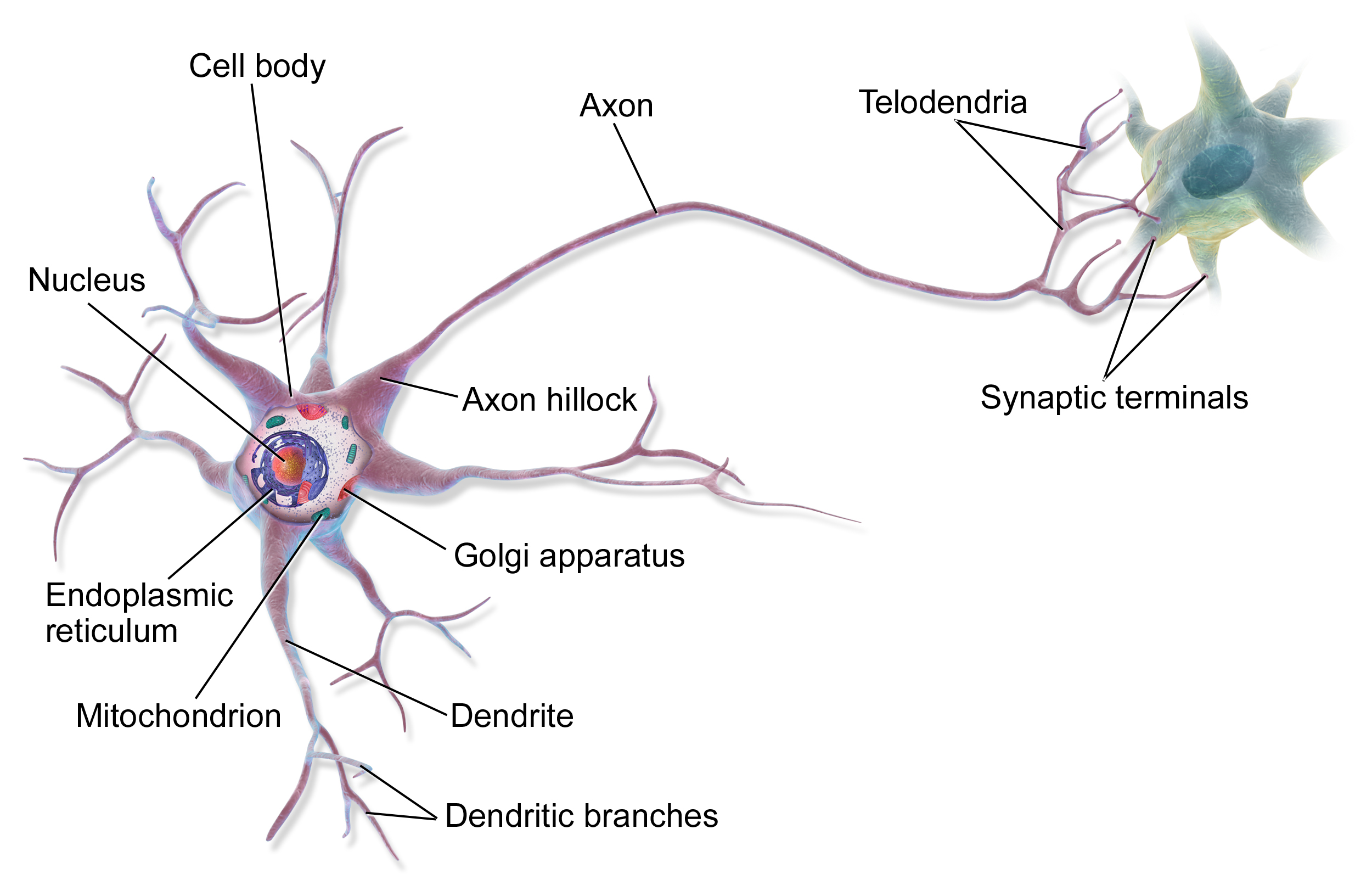
Neuron - Label - Labelled diagram - Wordwall Neuron - Label. Share Share by Aspillane. Y12 Y13 Psychology. Like. Edit Content. Embed. More. Leaderboard. Show more Show less . This leaderboard is currently private. Click Share to make it public. This leaderboard has been disabled by the resource owner. This leaderboard is disabled as your options are different to the resource owner. ... Label Neuron Anatomy Printout - EnchantedLearning.com Read the definitions, then label the neuron diagram below. axon - the long extension of a neuron that carries nerve impulses away from the body of the cell. cell body - the cell body of the neuron; it contains the nucleus (also called the soma) dendrites - the branching structure of a neuron that receives messages (attached to the cell body) Neuron Label - The Biology Corner Neuron Label. Publisher: Biologycorner.com; This work is licensed under a Creative Commons Attribution-NonCommercial 3.0 Unported License. This picture of the neuron is unlabeled, write in the labels to test your knowledge of the anatomy of a neuron. Neuron Label. ... An Easy Guide to Neuron Anatomy with Diagrams - SimplyPsychology.org Neurons, also known as nerve cells, are essentially the cells that make up the brain and the nervous system. Neurons do not touch each other, but where one neuron comes close to another neuron, a synapse is formed between the two. According to new research, the human brain contains around 86 billons neurons (Herculano-Houzel, 2009).
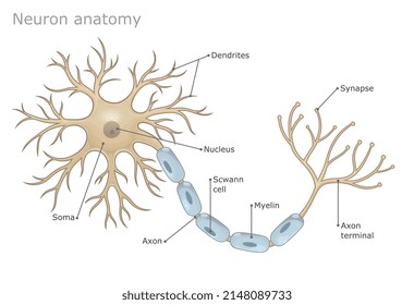
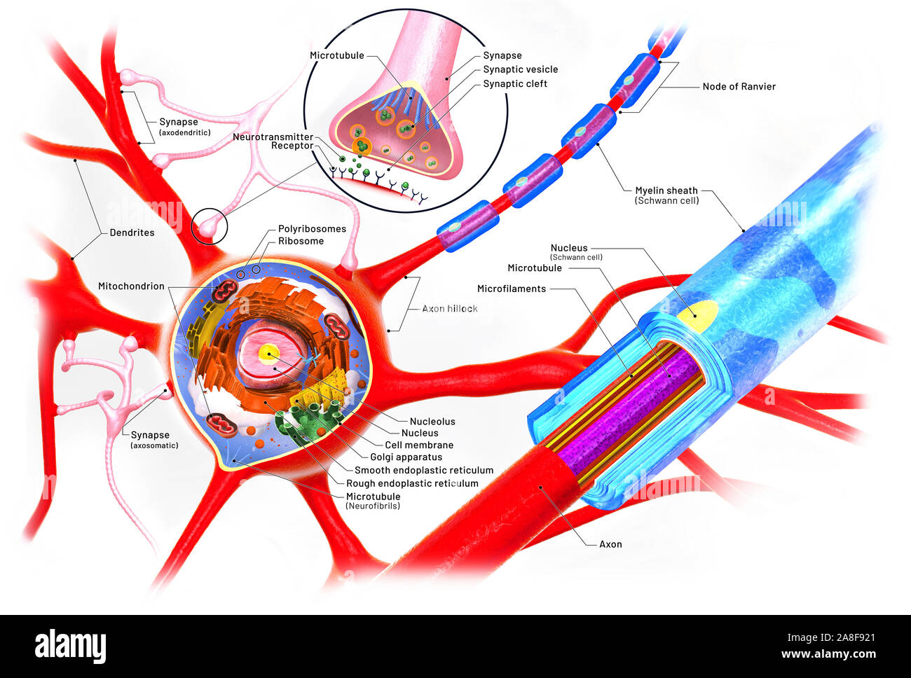
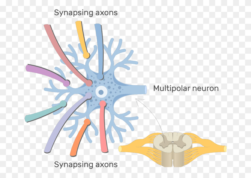
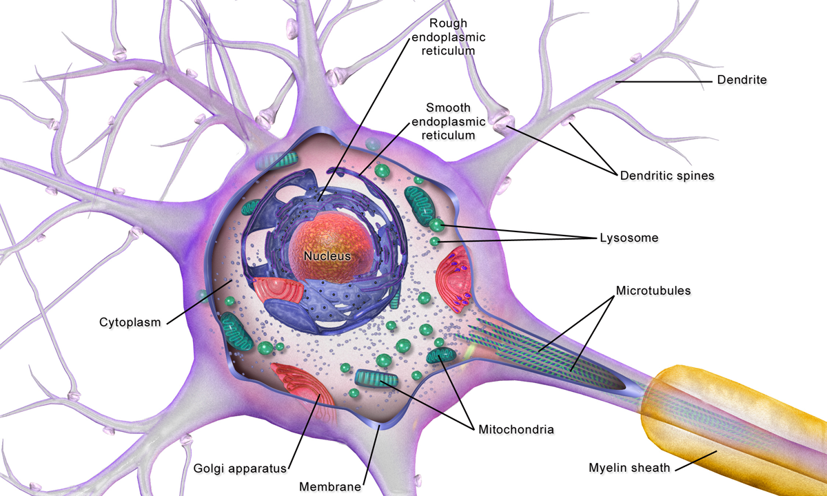
Types of Neurons: Parts, Structure, and Function - Verywell Health Summary. Neurons are responsible for transmitting signals throughout the body, a process that allows us to move and exist in the world around us. Different types of neurons include sensory, motor, and interneurons, as well as structurally-based neurons, which include unipolar, multipolar, bipolar, and pseudo-unipolar neurons. Neuron Diagram & Types | Ask A Biologist - Arizona State University Neurons pass messages to each other using a special type of electrical signal. Some of these signals bring information to the brain from outside of your body, such as the things you see, hear, and smell. Other signals are instructions for your organs, glands and muscles. Neurons receive these signals from neighbor neurons through their dendrites. Neuron under Microscope with Labeled Diagram - AnatomyLearner But, first, let's try to identify the following features from a neuron with the help of a labelled diagram. Cell body or perikaryon of a neuron Nucleus, cytoplasm, the plasma membrane of a neuron Nissl bodies in the cell body of a neuron An initial segment of axon and axon hillock Dendrites and axons of a neuron Axolemma and myelin sheath A Guide to Understand Neuron with Neuron Diagram To properly understand the coordination between the brain and the body, the students must learn about the neurons. They can use neuron-labeled diagrams while learning the complex structure of neurons. Creating a neuron-labeled image by hand can be difficult. The students must use the EdrawMax Online tool to make a high-quality neuron diagram . 2.
Neuron Label Diagram | Quizlet
Neuron Labeling - The Biology Corner This labeling activity was created for remote learning during the 2020 pandemic. Students in anatomy and physiology spent this year learning remotely, which required some adjustments in how material was presented. In the past, students would label a nerve cell and color neuroglia cells using paper handouts to learn the structures of the neuron ...
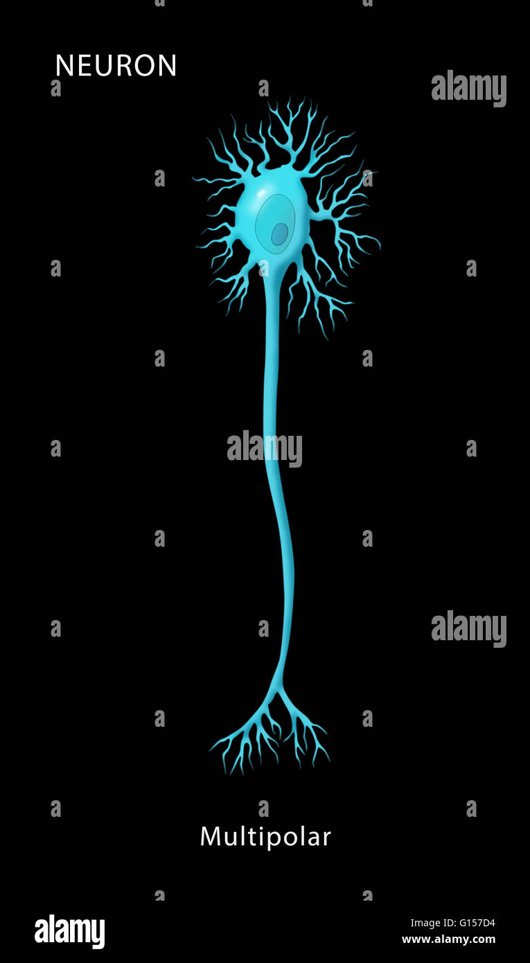
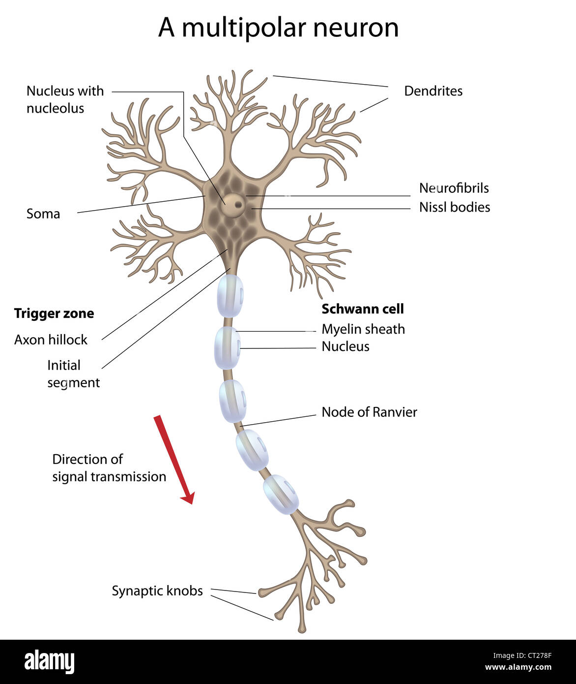
Labeled illustration of a multipolar neuron, one of the four ...
Neuron Label Flashcards | Quizlet Neuron Label Flashcards | Quizlet Neuron Label 5.0 (2 reviews) Term 1 / 9 Dendrites Click the card to flip 👆 Definition 1 / 9 ... Click the card to flip 👆 Flashcards Learn Test Match Created by meghangrossman Terms in this set (9) Dendrites Cell Body Nucleus of a neuron H Astrocyte Nodes of ranvier Axon Microglial cell Oligodendrocyte
File:Derived Neuron schema with no labels.svg - Wikimedia Commons
Label a Neuron Quiz - PurposeGames.com This is an online quiz called Label a Neuron. There is a printable worksheet available for download here so you can take the quiz with pen and paper. From the quiz author. Title says it all. Your Skills & Rank. Total Points. 0. Get started! Today's Rank--0. Today 's Points. One of us! Game Points. 10.
Nervous System - Label the Neuron
Labeled Neuron Diagram | Science Trends Neurons are the cells that are responsible for receiving sensory input from the outside world, sending motor commands to move parts of the body, forming memories in the brain, and more. Virtually every aspect of our experiential life and engagement with the world is mediated by the activity of neurons.

Identify the labels of the diagram.
Nerve Cell (Neuron) Labeling Page - Exploring Nature Nerve Cell (Neuron) Labeling Page. Higher Resolution PDF for Printing. Click Here. Use Teacher Login to show answer keys or other teacher-only items. Link to More About this Part of the Human Body. Click Here. Citing Research References. When you research information you must cite the reference. Citing for websites is different from citing from ...
564 Neuron labeled Images, Stock Photos & Vectors | Shutterstock
Nervous System - Label the Neuron - TheInspiredInstructor.com Name: Choose the correct names for the parts of the neuron. (1) (2) (3) (4) (5) (6) This neuron part receives messages from other neurons. (7) This neuron part sends on messages to other neurons. (8) This neuron part gives messages to muscle tissue. (9) This neuron part processes incoming messages.

Draw a labeled diagram of a neuron
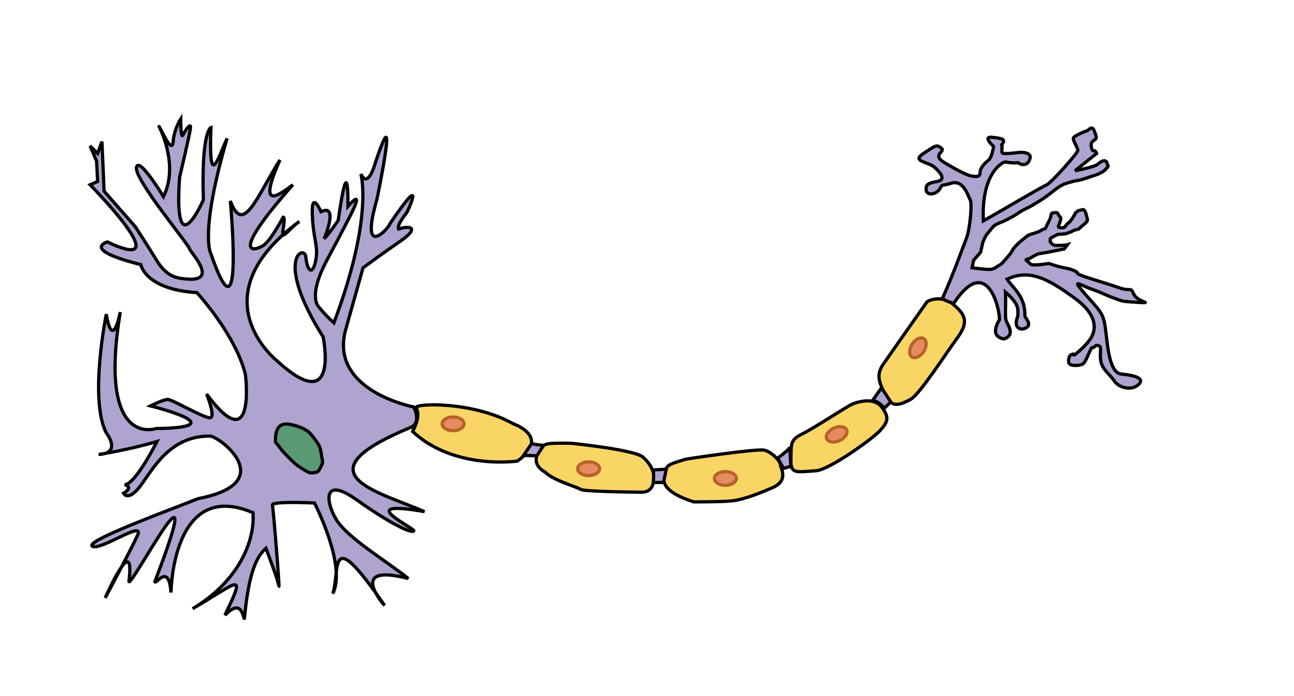
What Is a Neuron? - Definition, Structure, Parts and Function - BYJUS Neurons are the structural and functional unit of the nervous system. All neurons have three different parts - dendrites, cell body and axon. The neuron structure is specially adapted to carry messages over large distances in the body quickly in the form of electrical signals. What are sensory neurons and motor neurons?

The self-reconstructed 2.6 million rat cells without labels ...
Neuron Label Worksheets - K12 Workbook Displaying all worksheets related to - Neuron Label. Worksheets are Neuron anatomy activity, Work for classes 1 and 2, Neurotransmission fact, Neuron labeling work answers, Being brainy activity pack, Levels of organization foldable, Chapter 12 central nervous system, Brain anatomy. *Click on Open button to open and print to worksheet. 1.
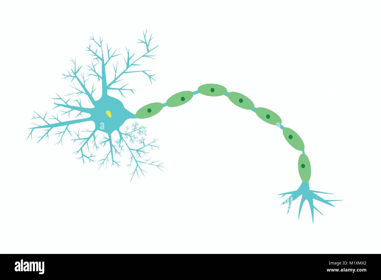
Illustration of structure of neuron, vector of schematic ...
Neuron Models - San Diego Mesa College Neuron Models. Click on a photo for a larger view of the model. Click on Label for the labeled model. Back to Nervous System
fDLC accurately predicts neuron labels of synthetic worms (A ...
Neuron Label Activity - Name each part of the neuron Explain the ... Name each part of the neuron Explain the function of each part. Dendrites Receive messages from other cells. They look like trees, so their "branches" stem on each side of the neuron so that it can detect the information. Cell Body The cell body is the bulky part of the neuron because that's where everything is held.
Labeled Neuron Diagram | Science Trends
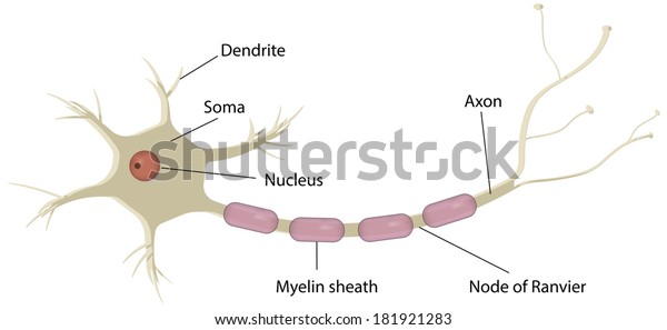
Neuron Diagram Labeled | EdrawMax Template It is an effective form of self-assessment, enabling students to check their understanding. In the following diagram, we have illustrated the important parts of the Neuron. In the following Neuron labeled diagram, we have dendrite, cell body, axon, myelin sheath, Schwann cell, a node of Ranvier, axon terminal, and nucleus.

Motor Neuron (labelled), illustration - Stock Image - C043 ...
A Labelled Diagram Of Neuron with Detailed Explanations - BYJUS They are found in the brain, spinal cord and the peripheral nerves. A neuron is also known as the nerve cell. The structure of a neuron varies with their shape and size and it mainly depends upon their functions and their location. Neurons are the structural and functional units of the nervous system. A group of neurons forms a nerve.

Labeled Neuron Diagram | Science Trends
Neuron Markers - BioLegend Neurons have highly compartmentalized structures that are generally classified into soma (cell body), axon, dendrite, axonal terminal, and synapse. BioLegend offers antibodies against markers that are expressed in each structural unit of a neuron, and allow their identification using applications such as microscopy, including immunohistochemistry (IHC) or immunocytochemistry (ICC).
Parts Of The Neuron Labeled, HD Png Download - kindpng
Diagram Quiz on Neuron Structure and Function (Labeling Quiz) Diagram Quiz on DNA replication. 1. Identify the cell type in the above figure. 2. In the figure, labeled '1' receives impulses from adjacent neuron. It is called the. 3. In the figure, labeled '2' is the short filaments from the cell body that carries impulses from dendrites to the cell body which is the. 4.
Nervous System Histology Illustrations - Neuron (labels ...
Il 407 R00 Nerve Cross Section W Labels - Sensory Neuron ...
Blausen 0657 - Multipolar neuron - English labels | AnatomyTOOL
Neuron to label hi-res stock photography and images - Alamy

File:Neuron-no labels.test.PNG - Wikimedia Commons
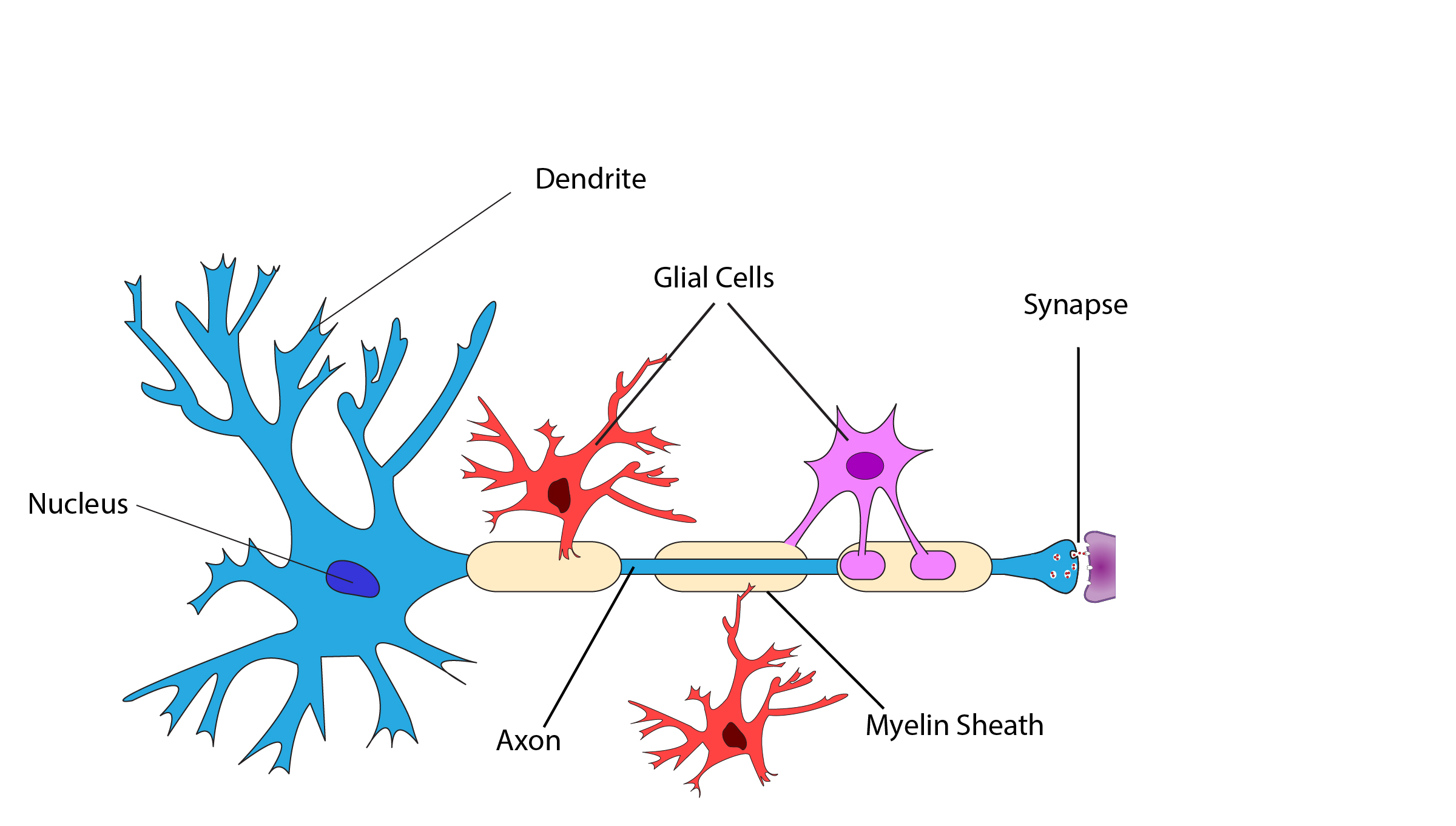
Fig 3 Neuron with labels dad remake - Exploring Medicine

neuron - BIOLOGY4ISC

CaMPARI2 photoconversion in vivo labels neurons based on ...

Servier - Drawing Neuron - no labels | AnatomyTOOL
Neuron Clipart Generic Labeled Multipolar Neuron Diagram ...
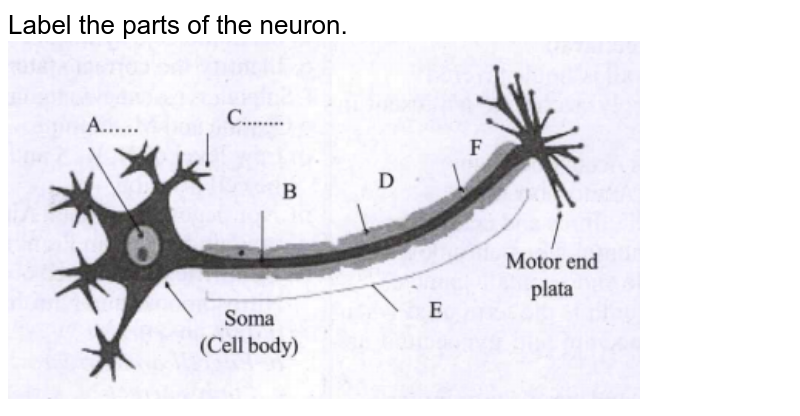
Label the parts of the neuron.

Label the parts of a typical neuron (1, 2, 3, 4, 5, 6) shown ...
Vektor Stok Nerve Cell Neuron Labeled Diagram (Tanpa Royalti ...

Labeling a Neuron Quiz

Ilustrasi Stok Motor Neuron Detailed Accurate Labeled ...

What Is a Neuron? Diagrams, Types, Function, and More

Labeled Parts of a Neuron stock illustration. Illustration of ...

Posts about neuron on BiologyWedsComputer | Neurons, Neuron ...
Blausen - Neuron cell body - English labels | AnatomyTOOL
Label A Neuron - Free Transparent PNG Clipart Images Download
Neuron to label hi-res stock photography and images - Alamy

Solved Label the structures below to explore the anatomy of ...
Motor neuron, labeled Stock Photo - Alamy

Label the parts of a typical neuron (1, 2, 3, 4, 5, 6) shown ...
Post a Comment for "38 neuron labels"