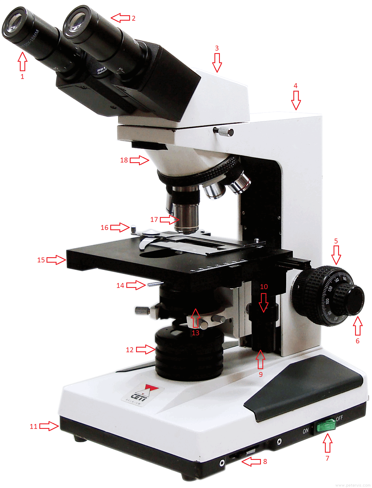
43 microscope diagram with labels
ch 8 mastering biology Flashcards | Quizlet Looking through a light microscope at a dividing cell, you see two separate groups of chromosomes on opposite ends of the cell. ... Drag the labels onto the diagram to identify the various chromosome structures. a. pair of homologous chromosomes b. centromere c. sister chromatides. Meiosis Animation Microscope Objective Lens | Products | Leica Microsystems The objective lens is a critical part of the microscope optics. The microscope objective is positioned near the sample, specimen, or object being observed. It has a very important role in imaging, as it forms the first magnified image of the sample. The numerical aperture (NA) of the objective indicates its ability to gather light and largely determines the microscope’s resolution, the ...
A Study of the Microscope and its Functions With a Labeled Diagram ... A Study of the Microscope and its Functions With a Labeled Diagram, To better understand the structure and function of a microscope, we need to take a look at the labeled microscope diagrams of the compound and electron microscope. These diagrams clearly explain the functioning of the microscopes along with their respective parts.

Microscope diagram with labels
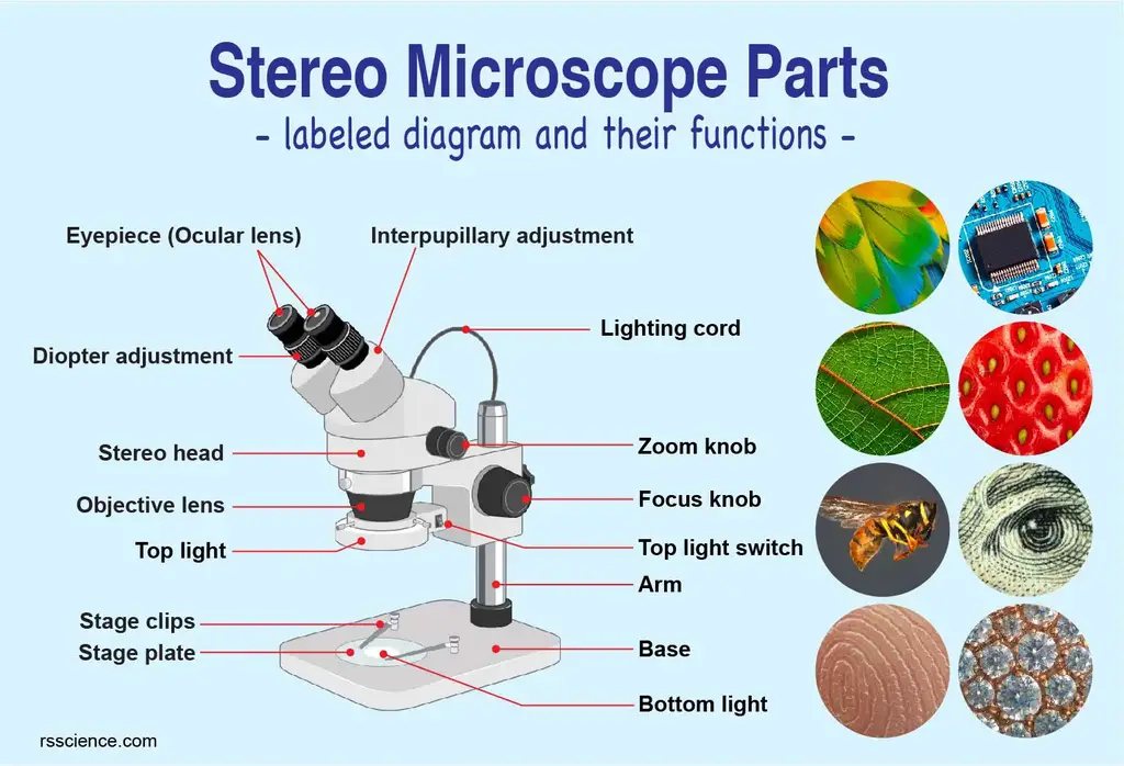
Parts of a microscope with functions and labeled diagram - Microbe Notes Parts of a microscope with functions and labeled diagram, April 19, 2022 by Faith Mokobi, Having been constructed in the 16th Century, Microscopes have revolutionalized science with their ability to magnify small objects such as microbial cells, producing images with definitive structures that are identifiable and characterizable. Microscope Labeling - The Biology Corner Microscope Labeling. This simple worksheet pairs with a lesson on the light microscope, where beginning biology students learn the parts of the light microscope and the steps needed to focus a slide under high power. The labeling worksheet could be used as a quiz or as part of direct instruction where students label the microscope as you go ... Parts of Stereo Microscope (Dissecting microscope) – labeled diagram ... Labeled part diagram of a stereo microscope Major structural parts of a stereo microscope. There are three major structural parts of a stereo microscope. The viewing Head includes the upper part of the microscope, which houses the most critical optical components, including the eyepiece, objective lens, and light source of the microscope.
Microscope diagram with labels. The Parts of a Microscope (Labeled) Printable - TeacherVision The Parts of a Microscope (Labeled) Printable. Worksheets. Science. The Parts of a Microscope (Labeled) Printable. Download. Add to Favorites. Share. This diagram labels and explains the function of each part of a microscope. Use this printable as a handout or transparency to help prepare students for working with laboratory equipment. Drawing Of A Microscope And Label - Warehouse of Ideas Each microscope layout (both blank and the version with answers) are available as pdf downloads. These labeled microscope diagrams and the functions of its various parts, attempt to simplify the microscope for you. Source: microbenotes.com, Drag and drop the text labels onto the microscope diagram. Microscope Poster - Diagram with Labels | Teach Starter A poster containing a diagram with labels showing the key parts of a microscope. In Science it is important that students know how to use a variety of tools when conducting scientific experiments and inquiry. This poster focuses on the microscope and highlights its key parts. Print on tabloid paper to display around your school's science lab ... PDF Label parts of the Microscope: Answers Label parts of the Microscope: Answers Coarse Focus Fine Focus Eyepiece Arm Rack Stop Stage Clip . Created Date: 20150715115425Z ...
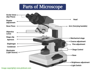
Parts of the Microscope with Labeling (also Free Printouts) Parts of the Microscope with Labeling (also Free Printouts) By Editorial Team March 7, 2022, A microscope is one of the invaluable tools in the laboratory setting. It is used to observe things that cannot be seen by the naked eye. Table of Contents, 1. Eyepiece, 2. Body tube/Head, 3. Turret/Nose piece, 4. Objective lenses, 5. Looking at the Structure of Cells in the Microscope A typical animal cell is 10–20 μm in diameter, which is about one-fifth the size of the smallest particle visible to the naked eye. It was not until good light microscopes became available in the early part of the nineteenth century that all plant and animal tissues were discovered to be aggregates of individual cells. This discovery, proposed as the cell doctrine by Schleiden and … The Cell - ScienceQuiz.net The diagram shows a group of onion cells. The parts labelled A, B and C respectively are? A = cell wall, B = cytoplasm, ... The diagram shows a plant cell as seen under a microscope. Two of the labels are incorrect. What are they?? Vacuole and chloroplast? Vacuole and cytoplasm? Nucleus and chloroplast? Microscope Parts and Functions A standard microscope has three, four, or five objective lenses that range in power from 4X to 100X. When focusing the microscope, be careful that the objective lens doesn't touch the slide, as it could break the slide and destroy the specimen. Specimen or slide: The specimen is the object being examined.
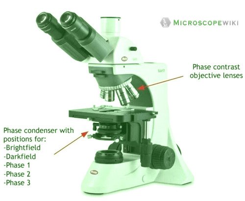
Confocal Microscopy - an overview | ScienceDirect Topics A standards document, which describes confocal microscopy and its influence quantities, has recently completed an ISO ballot as a final draft international standard (ISO FDIS 25178-607, 2018). A schematic diagram of a typical confocal microscope is shown in Fig. 15.1 (ASME B46-2009, 2010; Weller et al., 2012). Compound Microscope Parts, Functions, and Labeled Diagram Compound Microscope Parts, Functions, and Labeled Diagram, Parts of a Compound Microscope, Each part of the compound microscope serves its own unique function, with each being important to the function of the scope as a whole. Microscope Labeled Pictures, Images and Stock Photos photosynthesis. Diagram of the process of photosynthesis, showing the light reactions and the Calvin cycle. photosynthesis by absorbing water, light from the sun, and carbon dioxide from the atmosphere and converting it to sugars and oxygen. Light reactions occur in the thylakoid. Calvin Cycle occurs in the stoma. Neutrophil vector illustration. Label the microscope — Science Learning Hub 8.6.2018 · All microscopes share features in common. In this interactive, you can label the different parts of a microscope. Use this with the Microscope parts activity to help students identify and label the main parts of a microscope and then describe their functions.. Drag and drop the text labels onto the microscope diagram. If you want to redo an answer, click on the …
Labeling the Parts of the Microscope | Microscope World Resources
Microscope Diagram Labeled, Unlabeled and Blank | Parts of a Microscope ... FREE Microscope Parts Worksheet - This free microscope diagram printable is perfect for reviewing parts of microscope before your life science students or biology classes take out the microscopes for their next lab. Happy #teaching! #stem #freeprintable #biology, H, Help Teaching, Free Printable Worksheets,
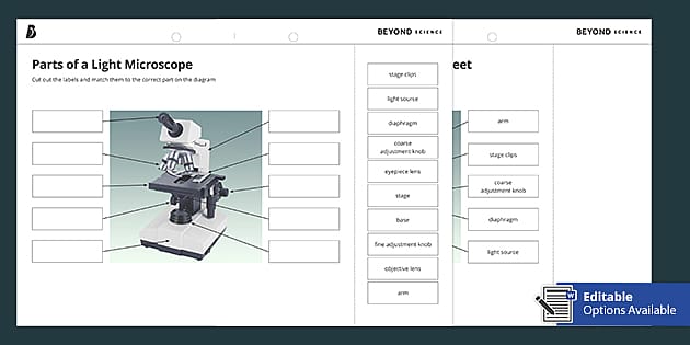
Parts of a Light Microscope Cut and Stick Worksheet - Twinkl
Labeling the Parts of the Microscope | Microscope World Resources Labeling the Parts of the Microscope, This activity has been designed for use in homes and schools. Each microscope layout (both blank and the version with answers) are available as PDF downloads. You can view a more in-depth review of each part of the microscope here. Download the Label the Parts of the Microscope PDF printable version here.

Microscopy: History, Types of Microscope, Diagram - Embibe
Microscope Diagram Labeled, Unlabeled and Blank | Parts of a Microscope ... Microscope Diagram Labeled, Unlabeled and Blank | Parts of a Microscope, Print a microscope diagram, microscope worksheet, or practice microscope quiz in order to learn all the parts of a microscope. Tim's Printables, 38k followers, More information, Microscope Diagram Unlabeled, Find this Pin and more on Chemistry by Ginger Dwyer.

Compound Microscope Parts – Labeled Diagram and their ...
Parts of a Microscope Labeling Activity - Storyboard That Create a poster that labels the parts of a microscope and includes descriptions of what each part does. Click "Start Assignment". Use a landscape poster layout (large or small). Search for a diagram of a microscope. Using arrows and textables label each part of the microscope and describe its function. Copy This Storyboard*, More options,

Ecology Test Day Microscope Introduction Day (Test day ...
Microscope Labeling - The Biology Corner Students label the parts of the microscope in this photo of a basic laboratory light microscope. Can be used for practice or as a quiz. ... 20. A microscope has an ocular objective of 10x and a high power objective of 50x, what is the microscope's total magnification? _____
Microscope Diagram Parts - ClipArt Best
Microscope Parts, Function, & Labeled Diagram - slidingmotion Microscope parts labeled diagram gives us all the information about its parts and their position in the microscope. Microscope Parts Labeled Diagram, The principle of the Microscope gives you an exact reason to use it. It works on the 3 principles. Magnification, Resolving Power, Numerical Aperture. Parts of Microscope, Head, Base, Arm,

This is a common compound microscope. Label its parts from A ...
Fluorescence Resonance Energy Transfer (FRET) Microscopy Presented in Figure 3 is a Jablonski diagram illustrating the coupled transitions involved between the donor emission and acceptor absorbance in fluorescence resonance energy transfer. Absorption and emission transitions are represented by straight vertical arrows (green and red, respectively), while vibrational relaxation is indicated by wavy yellow arrows.

Draw a neat labelled diagram of a compound microscope class ...
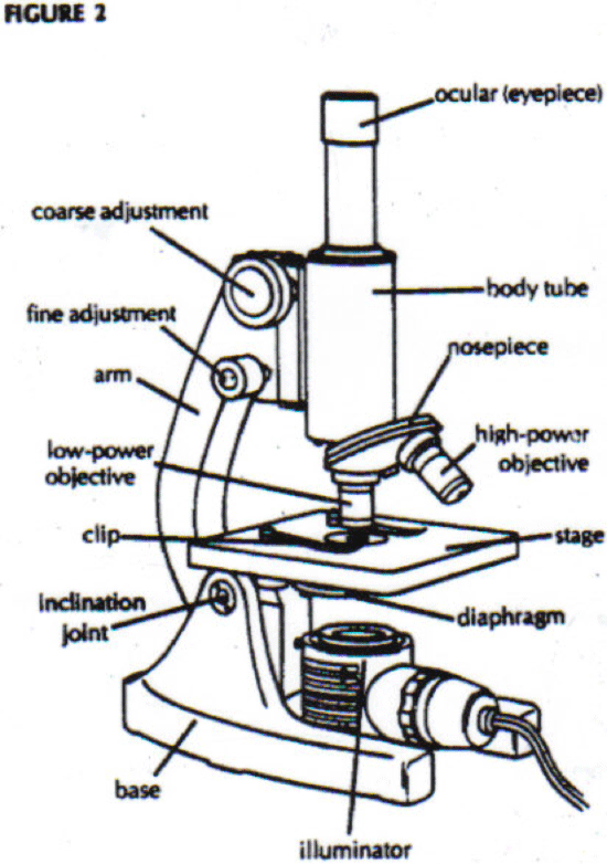
Labelled Diagram of Compound Microscope The below mentioned article provides a labelled diagram of compound microscope. Part # 1. The Stand: The stand is made up of a heavy foot which carries a curved inclinable limb or arm bearing the body tube. The foot is generally horse shoe-shaped structure (Fig. 2) which rests on table top or any other surface on which the microscope in kept.

GCSE Optical microscope labelling Diagram | Quizlet
Compound Microscope- Definition, Labeled Diagram, Principle, Parts, Uses The optical microscope often referred to as the light microscope, is a type of microscope that uses visible light and a system of lenses to magnify images of small subjects. There are two basic types of optical microscopes: Simple microscopes. Compound microscopes. The term "compound" in compound microscopes refers to the microscope having ...

Compound Microscope: Parts of Compound Microscope
Compound Microscope Parts - Labeled Diagram and their Functions Labeled diagram of a compound microscope, Major structural parts of a compound microscope, There are three major structural parts of a compound microscope. The head includes the upper part of the microscope, which houses the most critical optical components, and the eyepiece tube of the microscope.
MICROBIO 16 Parts of a Compound Microscope with Diagram and ...
Label the Microscope Diagram | Download Scientific Diagram - ResearchGate Download scientific diagram | Label the Microscope Diagram from publication: Laboratory Exercises in Microbiology: Discovering the Unseen World through Hands-on Investigation | Microbiology ...

List: Parts of a Microscope and their Function | Pathwooded
16 Parts of a Compound Microscope: Diagrams and Video Once you have an understanding of the parts of the microscope it will be much easier to navigate around and begin observing your specimen, which is the fun part! The 16 core parts of a compound microscope are: Head (Body) Arm. Base. Eyepiece. Eyepiece tube.

Microscope Labeling Activity - SMART Board Activity - Interactive Review
(PDF) Introduction to Microscopy - ResearchGate 8.11.2017 · Introduction to Microscopy, its different types in optical and electron based microscopy. Also presentation involved working principles of Optical, SEM & TEM microscope with their components ...

Simple Microscope Definition, Magnification, Parts And Uses
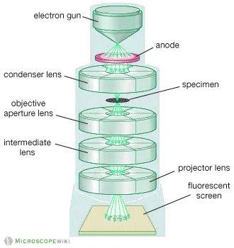
Electron microscope - Wikipedia An electron microscope is a microscope that uses a beam of accelerated electrons as a source of illumination. As the wavelength of an electron can be up to 100,000 times shorter than that of visible light photons, electron microscopes have a higher resolving power than light microscopes and can reveal the structure of smaller objects.. Electron microscopes use shaped magnetic …
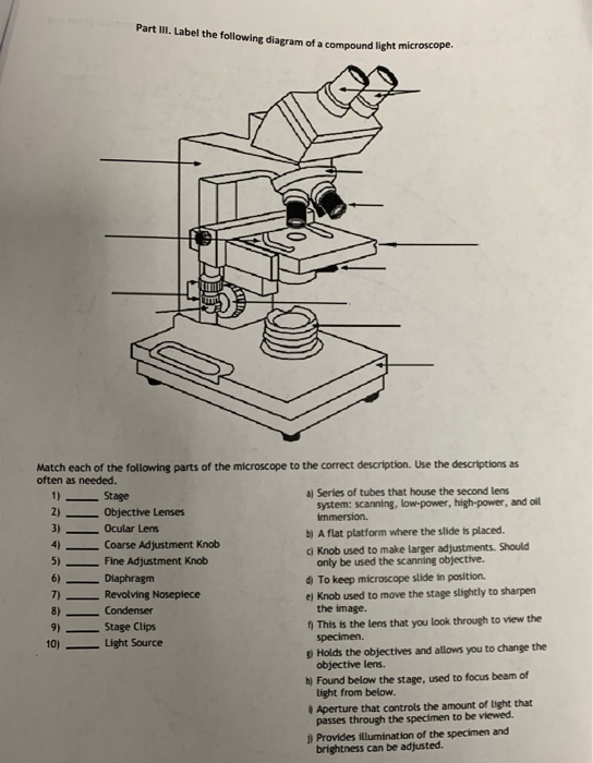
Solved Part III. Label the following diagram of a compound ...
Microscope, Microscope Parts, Labeled Diagram, and Functions Microscope, Microscope Parts, Labeled Diagram, and Functions, What is Microscope? A microscope is a laboratory instrument used to examine objects that are too small to be seen by the naked eye. It is derived from Ancient Greek words and composed of mikrós, "small" and skopeîn,"to look" or "see".
biology labeled microscope diagram - Clip Art Library
Labeled Microscope and Basics of Life Diagram | Quizlet PLAY. A microscope is an instrument widely to magnify and resolve the image of an object that is otherwise invisible to naked eye. For resolving the details of objects, which otherwise cannot be achieved by naked eye, a microscope is used. This set of flash cards will help the student to identify the different parts and function of the microscope.

Produk Microscope | UD Berkah Abadi
Simple Microscope - Diagram (Parts labelled), Principle, Formula and Uses Parts of a Simple Microscope, A simple microscope consists of, Optical parts, Mechanical parts, Labeled Diagram of simple microscope parts, Optical parts, The optical parts of a simple microscope include, Lens, Mirror, Eyepiece, Lens, A simple microscope uses biconvex lens to magnify the image of a specimen under focus.
DC5-420TH Stereo Zoom Microscope from National Optical ...
Microscope Types (with labeled diagrams) and Functions Simple microscope labeled diagram, Simple microscope functions, It is used in industrial applications like: Watchmakers to assemble watches, Cloth industry to count the number of threads or fibers in a cloth, Jewelers to examine the finer parts of jewelry, Miniature artists to examine and build their work,
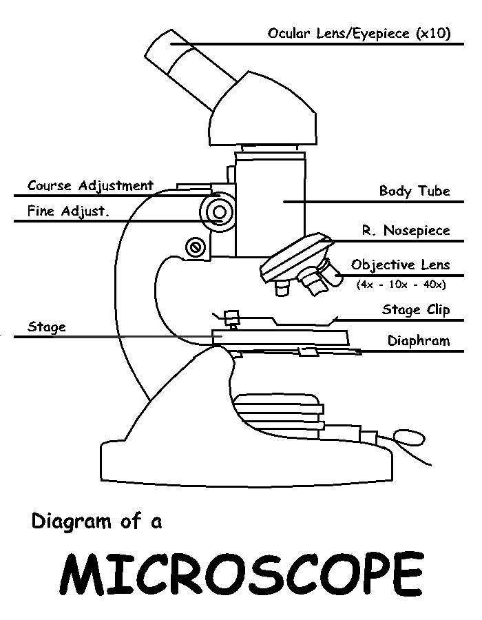
Diagram of a Microscope by ScienceDoodles on DeviantArt
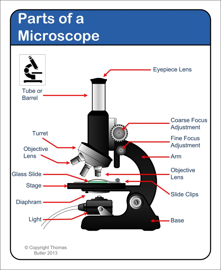
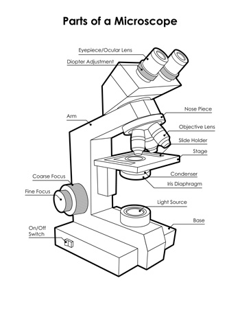
Label Microscope Diagram - EnchantedLearning.com arm - this attaches the eyepiece and body tube to the base. base - this supports the microscope. body tube - the tube that supports the eyepiece. coarse focus adjustment - a knob that makes large adjustments to the focus. diaphragm - an adjustable opening under the stage, allowing different amounts of light onto the stage.

draw a well label diagram of microscope - Brainly.in
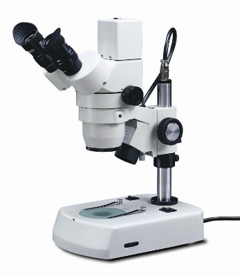
Parts of Stereo Microscope (Dissecting microscope) – labeled diagram ... Labeled part diagram of a stereo microscope Major structural parts of a stereo microscope. There are three major structural parts of a stereo microscope. The viewing Head includes the upper part of the microscope, which houses the most critical optical components, including the eyepiece, objective lens, and light source of the microscope.
Electron Microscope Principle, Uses, Types and Images ...
Microscope Labeling - The Biology Corner Microscope Labeling. This simple worksheet pairs with a lesson on the light microscope, where beginning biology students learn the parts of the light microscope and the steps needed to focus a slide under high power. The labeling worksheet could be used as a quiz or as part of direct instruction where students label the microscope as you go ...

Labeling the Parts of the Microscope | Microscope World Resources
Parts of a microscope with functions and labeled diagram - Microbe Notes Parts of a microscope with functions and labeled diagram, April 19, 2022 by Faith Mokobi, Having been constructed in the 16th Century, Microscopes have revolutionalized science with their ability to magnify small objects such as microbial cells, producing images with definitive structures that are identifiable and characterizable.
Parts of Stereo Microscope (Dissecting microscope) – labeled ...

The Microscope

Diagram of a Compound Microscope

Draw a labelled diagram of a compound microscope.
Compound Microscope Parts, Functions, and Labeled Diagram ...

5 Important Types of Microscopes used in Biology (With Diagram)

Diagram of traveling microscope setup with implant cast and ...
Junior cert Labelling Microscope - Labelled diagram
Parts of Microscope, Microscope Labeled Diagram and Functions ...

Microscope, Microscope Parts, Labeled Diagram, and Functions

Label the microscope — Science Learning Hub

Microscope Maintenance Tips | Science supplies, Microscope ...

Light Microscope- Definition, Principle, Types, Parts ...

Can someone can send me diagram of this compound microscope ...
Labeled Microscope Diagram - Tim's Printables

Simple Microscope - Diagram (Parts labelled), Principle ...
Labelled Diagram of Microscope Parts

microscope drawing with label - Clip Art Library

Labeled Microscope Diagram | Microscope parts, Science fair ...

Label Microscope Diagram - EnchantedLearning.com
Microscope Types (with labeled diagrams) and Functions
Post a Comment for "43 microscope diagram with labels"